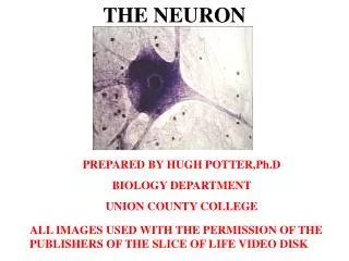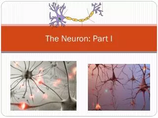
Session 4 The Neuron
PS111 Brain & Behaviour Module 1: Psychobiology. Session 4 The Neuron. What are neurons good for?. Why do more complex organisms need a nervous system?. In complex organisms, cells ... on the inside of the body are not in direct contact with the outside world

Session 4 The Neuron
E N D
Presentation Transcript
PS111 Brain & Behaviour Module 1: Psychobiology Session 4The Neuron
What are neurons good for? • Why do more complex organisms need a nervous system? • In complex organisms, cells ... on the inside of the body are not in direct contact with the outside world ... live in different ‘environments’ ... have become specialised • In order for the organisms to function, • cell activities must be co-ordinated
What are neurons good for? • Two systems to co-ordinate cell activities: a) Endocrine system: • specialised to secrete chemicals (‘hormones’) into the bloodstream • provide slow, overall co-ordination of cell activities b) Nervous system: • specialised to transmit electrical impulses between two or more cells • provide fast and precise co-ordination
What are neurons good for? Hi, Mike!
What are neurons good for? Hi, Mike!
What are neurons good for? • Neural impulses (‘signals’) provide constant & rapid communication between cells. • Signals from one (group of) cells change properties of receiving cell • => i.e., change the way the receiving cell ‘behaves’ In other words: • Neural impulses provide constant & rapid • control & adjustment of ongoing cell activities • QUESTIONS: • How are neural impulses generated? • How are they transmitted? • What is their function?
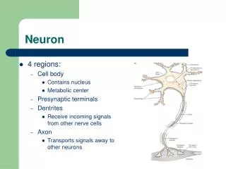
Neurons are special! 1. Function: • Generation & transmission of electrical impulses • Rapid • Over great distances • Point-to-point • Electrical impulses reach specific targets • Modifies activity of the target cells • Allows selective control of specific target structures • Electrical activity modulated by integrated input from other cells • ‘Input’ used to adjust ‘output’ • Combination & integration of signals from different sources • Structured communication
Neurons are special! Smooth muscle cell Skin cells Neural (pyramidal) cell Ovary cell Blood cells 2. Form & Size:
Neurons are special! 3. Special requirements: Virtually no possibility to store energy • Glucose (sugar) & oxygen must be constantly supplied • Without supply, neurons • stop working within seconds • die within minutes 4. Life span: Neurons do not divide (they develop from ‘neural stem cells’) • Neurogenesis virtually completed around 5 months after conception: • after this, dead neurons can not be replaced (mostly) • Neuron death part of normal brain development: • 20% to 80% of all neurons die during maturation because neurons are so special…
Glia Cells • Provide ‘protected environment’ for neurons to survive • Develop – like neurons – from neural stem cells • About 10 times as many glia as neurons, • on average 1/10 the size of a neuron
Glia Cells http://i.livescience.com/images/060105_astrocyte_02.jpg Xu, Pan, Yan, & Gan, NatNeuro,10, 549-551. http://www.nature.com/neuro/journal/v10/n5/images/nn1883-F2.jpg • Astrocytes: • star-shaped • physical & nutritional support for neu-rons (part of Blood-Brain-Barrier): • transport nutrients from blood vessels to neurons and • waste products away from neurons • hold neurons in place • Play a role in neural signal transmission as well! • Microglia: • small • mobile for defensive function: • produce chemicals that aid repair of damaged neurons • digest dead neurons (phagocytosis)
Glia Cells • Oligodendroglia: • large, flat branches • wrapped around axons • consist of fatty sub-stance (myelin) • insulating the axon Other types of glia exist, but will not be discussed here...
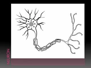
Neurons Dendrites Membrane Axon Hillock Axon terminals Axon Soma Neural Signal Transmission...
Positively charged ions Negatively charged ions • Neurons are not empty, and do not exist in a vacuum: • Thick chemical ‘soup’ of electrically charged particles • fills the neuron (‘intra-cellular fluid’) • surrounds the neuron (‘extra-cellular fluid’) • (now recall that the membrane has holes)
Electrical activity - Resting potential Cl 1. Basic principles: Concentration Gradient Electrical Gradient
Electrical activity - Resting potential • Ion concentrations differ between the inside and the outside of the cell: Na+Cl- K+ Na+Cl- + K+ Na+Cl- - - A- K+A- - (-70 mV) Na+Cl- K+ Na+Cl- + Concentration Gradient K+ Na+ Cl- A- K+ A- - Electrical Gradient 2. Ion gradients: • Electrical potential remains static => no electrical activity • Protein channels in cell membrane • allow ions to enter or leave the cell:
Electrical activity - Resting potential K+ Cl- 0 K+ Na+ Cl- K+ Na+Cl- A- Na+A- 0 • Active channels work against the equilibrium: Na+Cl- K+ Na+ Cl- Na+ Na+ Na+ + - (-70 mV) K+ Na+ Cl- A- K+ A- K+ K+ 2. Ion gradients: • If channels were passive ‘holes’, membrane would depolarise (electrical potential would disappear): • Again, there would be no electrical activity! 3. Sodium/potassium pump and membrane potential: • Neurons need energy just to maintain their resting potential!
Electrical activity – Signal Transmission • Based on movement of electrically charged particles (ions): • Ion-specific channels in cell membrane are GATES that canopen(they are not open all the time!) • either by chance or in response to stimulation • Positive or negative ions enter or leave the cell • Depolarisation: • Positive ions in, or negative ions out: • Inside less negative than usually • Hyperpolarisation: • Negative ions in, or positive ions out: • Inside more negative than usually
Electrical activity – Signal Transmission Electrotonic Action Potential Synaptic • Passive: Ions move inside the cell along electrical & concentra-tion gradients. • Some ions will get lost on their way: Signal decays over time
Electrical activity – Signal Transmission Electrotonic Action Potential Synaptic • Active (self-replicating, no decay): ions move locally through cell membrane. • Generated at axon hillock, moves down the axon towards ter-minal buttons
Electrical activity – Action Potential Electrical stimulation: K+ Cl- Na+ Na+ Na+ Na+ Na+ Na+ Na+ Na+ Na+ Na+ + Resting potential: Na+ Na+ Na+ Na+ Na+ Na+ Na+ Na+ Na+ Na+ K+ Na Na+ Na+ Cl- Na+ Na+ Na+ Na+ Na+ K+ Na+ Cl- A- A- + + - (-70 mV) K+ K+ K+ Cl- Na+ Na+ Na+ K+ Cl- K+ Cl- Na+ Na+ Na+ Na+ Na+ Na+ Na+ Na+ Na+ Na+ K+ Na Cl- Na+ Na+ Threshold: Na+ Na+ inflow: K+ Na Cl- Na+ Na+ Na+ Na+ K+ Na+Cl- A- A- K+ Na Cl- Na+ Na+ Na+ Na+ K+ Na+ Cl-A- A- (-70 mV) K+ Na+ Cl- A- A- Na+ K+ K+ (-50 mV) Na+ (-50 mV) K+ Cl- Na+ K+ K+ Na+ Na+ K+ K+ Na+ Na+ K+ Na Cl- Na+ Na+ Na+ Na+ K+ Na+ Cl- A- A- Na+ Na+ Na+ (+50 mV) Na+ Na+ K+ K+ Na+ Na+ Na+ Na+ Na+ 1. Voltage gated membrane channels: • Na+ channels open or close in response to electrical changes at the membrane • Sequence of events: 1. Membrane depolarised (inside less negative) 2. SomeNa+ channels open 3. Na+ ions enter the cell 4. Membrane depolarises further -- THRESHOLD? 5. All nearby Na+ channels open 6. Membrane fully depolarised (more positive on the in- than on the outside!)
Electrical activity – Action Potential Electrical stimulation Membrane depolarises Na+ inflow Na+ channels open 2. Threshold potential and the Hodgkin-Huxley cycle: • If membrane potential at axon hillock remains below threshold, resting potential returns • If membrane depolarises further: • more and more Na+ channels will open, • resulting in more and more depolarisation • If membrane potential at axon hilockreachesthreshold: • allNa+ channels in depolarised area open simultaneously, • generating an action potential
Electrical activity – Action Potential Na+channel close, K+channel open K+ OUT 3. Electrochemical processes during an AP • Threshold has been reached: • so many Na+ ions enter the cell that inside becomes more positive than outside (complete depolarisation) • Complete depolarisation causes • a) Closing of Na+ channels: • No more Na+ions enter cell • b) Opening of K+ channels: • K+ ions rush out of cell: • membrane repolarises Na+ channel open Na+ IN • K+ channels close when resting potential is restored • briefly, less K+ ions inside than outside cell: • membrane hyperpolarized (inside more negative than usual)
Electrical activity – Action Potential 4. Conduction of the action potential • Originates at axon hillock & travels down the axon • Each burst of depolarisation acts as a trigger, • opening Na+ channels in adjacent regions of the axon • Why does the action potential not travel backwards? • During hyperpolarisation, mem-brane more difficult to depo-larise • But adjacent part of axon (where AP has not yet occurred) easy to depolarise
Electrical activity – Action Potential 5. Properties of the action potential: • No decay: • always strong enough to depolarise adjacent membrane • ‘All-or-nothing’ phenomenon: • either generated or not • can not be generated with different intensities! • Discontinuous: • minimal time between subsequent APs: 2-5ms • Fast: • approx. 1-10 m/s • However, for some purposes, this might not be fast enough
Electrical activity – Action Potential 6. Saltatory conduction • In mammals, the axons of sensory and motor neurons are myelinated • Myelin insulates, preventing ion inflow and outflow • Electrical charges transported inside the axon • no need to produce an AP
Electrical activity – Action Potential 6. Saltatory conduction • In mammals, the axons of sensory and motor neurons are myelinated • Myelin insulates, preventing ion inflow and outflow • Electrical charges transported inside the axon • no need to produce an AP • Nodes of Ranvier: • gaps that interrupt insulation every 1-2 mm Node of Ranvier + + + + + + + + + + + + + + + + + + + + + + + + + + + + + + + + + + + + + + + + + Axon + + + + + + + + + + + + + + + + Myelin + + + + + + + + + + + + + + + + + + + + + + + + + + + + + + +
Electrical activity – Action Potential 6. Saltatory conduction • In mammals, the axons of sensory and motor neurons are myelinated • Myelin insulates, preventing ion inflow and outflow • Electrical charges transported inside the axon • no need to produce an AP • Nodes of Ranvier: • gaps that interrupt insulation every 1-2 mm + + + + + + + + + + + + + + + + + + + + + + + + + + + + + + + + + + + + + + + + + + + + + + + + + + + + + + + + + + + + + + + + + + + + + + + + + + + + + + + + + +
Electrical activity – Action Potential 7. Signal transmission and information: • Electrical impulses can not be modified! • How are different types of information ‘coded’? • Qualitative: by location • the place in the brain where the signal is received • (cf. last lecture) • Quantitative (how strong a stimulus is): by ‘firing rate’ • a strong input causes a neuron to send out APs in quicker succession Weak stimulus: Strong stimulus: voltage voltage time time
Electrotonic Action Potential Synaptic Signal Transmission (details in the next lecture…)
QUESTION TIME • 1. In the figure below, the number 3 indicates the • a) pons • b) thalamus • c) corpus callosum • d) limbic system • e) cerebellum 3 4 2 5 1