
PERIAPICAL DISEASE
E N D
Presentation Transcript
Classification of inflammatory processes of MFA. Periodontitis: etiology, pathogenesis, classification, clinical course, complications, prophylaxis. Odontogenic granuloma of the face: clinic, treatment. Detained and halfdetained teeth. Etiology, clinic, diagnostics, treatment, complications. Pericoronaritis. Odontogenic jaw periostitis: etiology, clinic, diagnostics, treatment, complications, prophylaxis.
PERIAPICAL DISEASE Classified as: • Acute Apical Periodonitis • Acute Apical Abscess • Chronic Apical Periodontitis (Diffuse, Suppurative Apical Periodontitis with sinus tract, Apical cyst) • Condensing Osteitis
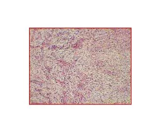
Definition • The fundamental lesion of chronic periapical inflammation is known as ´´chronic apicalperiodontitis´´ • While this designation is the preferred one, most dentists know it by the term ´´dental granuloma´´ • The lesion is not a granuloma at all because it is not composed of granulomatous chronic inflammation.
Classification • 1) Diffuse type: - small, recurrent amount of tissue damage - cellular infilltration with lymphocytes, plasma cells, phagocytic mononuclear cells, fibroblasts which produce granulation tissues for repair of damaged area GRANULOMA: formation of large nodule of granulation tissue that is slowly increase in size Resorption of hard tissue, granulation tissue around apex (outlined by capsule of fibrous tissue)
2)Chronic suppurative periodontitis -central cavity which is accompanied with fistula and stroma - its known as chronic apical abscess ( chronic alveolar abscess) • 3) Apical cyst - true cyst: pathologic cavity which contain fluid or semi-fluid substance that is lined by epithelium and surrounded by connective tissue capsule
Case 1,fig.1a 21-years old woman-non successful endodontic treatment tooth N.22,apical clear radiolucency confirming an established lesion bigger than 3mm,it shows features of lamina dura disruption and bone structural changes Case 1,fig.1b Measurement of the tooth canal length
Case 1,fig.1c Final endodontic treatment Foredent and gutapercha Case 1,fig.1d 5 months after the endodontic treatment without any surgical procedure,intraoral x-ray shows chronic apical periodontitis, partial restitution of the periapical region
Case 2,fig.2a Orthopantogram image,unsuccessful endodontic treatment d.N.22, Cystis radicularis D.N.22
Case 2,fig.2c 3months after the therapy-Cystectomio sec.PARTSCH II. et resectio apicis dentis N.22 Retrograde root canal endodontic therapy with amalgam Egalisatio,suturae Case 2,fig.2b Intraoral image D.22-Cystis radicularis processus alveolaris maxillae reg.frontalis purulenta
Fig.B Granuloma periapicalis and infection transmission paths
Chronic apical periodontitis. Extensive tissue destruction in the periapical region of a mandibular first molar occurred as a result of pulpal necrosis. Lack of symptoms together with presence of a radiographic lesion is diagnostic.
Periapical radiolucencies associated with mandibular incisors. These teeth were vital, and a diagnosis of cemental dysplasia was made.
Periodontitis chronica circumscripta d.14 Periodontitis chronica circumscripta d.41
CLINICAL FEATURES • HYPERSENSITIVE TOOTH UPON BITING OR PERCUSSION • NEGATIVE RESULTS IN BOTH ELECTRIC OR THERMAL STIMULI • BEING ACUTE IN NATURE, ON RADIOGRAPH THERE IS MILD THICKENING OF THE APICAL PERIODONTAL LIGAMENT SPACE. • IN CASES OF RECURRING CHRONIC EVENTS, PERIAPICAL CHANGES (LUCENCIES) MAYBE SEEN (PERIAPICAL GRANULOMA)
PERIAPICAL GRANULOMA • IN CASES OF LOW GRADE BUT CHRONIC INFLAMMATION AT THE APEX OF A NON VITAL TOOTH GRANULOMA IS USED ON AGAINST THE TERM ABSCESS WHICH IS OF ACUTE IN NATURE.
TREATMENT • DRAINAGE ESTABLISHMENT WITHIN THE TOOTH ITSELF OR ON THE SURROUNDING SOFT TISSUES • ANTIBIOTIC THERAPY • SKILLED AND THOUGHTFUL MANAGEMENT MUST BE EMPLOYED SINCE ANY DELAY MAY CAUSE ANY LETHAL CONSEQUENCE.
COMPLICATIONS • PUS MAY DRAIN ON NATURALLY OCCURING DRAINS TERMED AS FISTULAS OR SINUS TRACTS WHICH MAY BE SEEN ON SKIN OR ON THE PALATE • IF THERE IS NO DRAIN MADE CELLULITIS ENSUES AFTER THE PUS BUILDUP. IT IS AN ACUTE INFLAMMATORY SPREAD ON THE NEARBY SOFT TISSUES • ENZYMES ARE PRODUCED BY HIGHLY VIRULENT MICROORGANISMS PRESENT
COMPLICATIONS • BILATERAL SUBMANDIBULAR AND SUBLINGUAL SPACES ARE KNOWN AS “LUDWIG'S ANGINA” • FATALITIES USUALLY RESULTS FROM BACTEREMIA FROM INFECTION SPREADING INTO THE MAJOR BLOOD VESSELS OR THROUGH A RETROGRADE SPREAD OF INFECTION INTO THE FACIAL EMISSARY VEINS INTO THE CAVERNOUS SINUS, CAVERNOUS SINUS THROMBOSIS
IMPACTED TEETH • An impacted tooth is one that is partially erupted or unerupted and will not eventually assume a normal arch relationship withother teeth and tissues.
Causes of Impacted Teeth • Role of civilization • Local causes of Impaction • Systemic causes of impaction
Local causes of Impaction • Lack of space in the dental arch for eruption; • The density of the overlying or surrounding bone; • Long continued chronic inflammation with resultant increase in the density of the overlying mucous membrane; • Premature loss of the primary teeth; • Acquired diseases, such as necrosis due to infection or abscesses, and inflammatory changes in the bone due to exanthematous diseases in children;
Impacted teeth occur in the following order • Mandibular third molars • Maxillary third molars • Maxillary cuspids • Mandibular bicuspids • Mandibular cuspids • Maxillary bicuspids • Maxillary central incisors • Maxillary lateral incisors • Maxillary or mandibular first molars are rarely impacted
Classification of impacted mandibular third molars • A. Relation of the tooth to the ramus of the mandible and the second molar; • B. Relative depth of the third molar in bone; • C. The position of the long axis of the impacted mandibular third molar in relation to the long axis of the second molar : vertical, horizontal, inverted, mesioangular, distoangular, buccoangular, linguangular.
Class1 • the space between the anterior part of the ascending ramus and the distal surface of the 2nd molar is sufficient to accommodate the mesiodistal diameter of the crown of the third molar.
Class2 • the space between the anterior part of the ascending ramus and distal surface of the 2nd molar is less than the mesiodistal diameter of the crown of the third molar (part of the tooth located within the ramus)