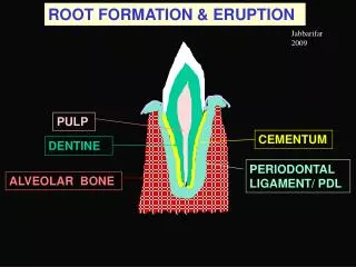
Pulp and Periapical Chapter 3
Pulp and Periapical Chapter 3 . Also notes from biopsy techniques. Teeth are non-vital. Condensing Osteitis.

Pulp and Periapical Chapter 3
E N D
Presentation Transcript
Pulp and Periapical Chapter 3 • Also notes from biopsy techniques
Condensing Osteitis • Two periapical films showing well defined radiopacity at apex of Mn 1st molar, exibits root tip absorption and loss of lamina dura and some widening of the PDL space. Both lesions are present on teeth with crown or extensive caries • Differential • Condensing osteitis--look for large carious lesion or crown (this is correct for previous 2 radiographs) • Idiopathic osteosclerosis (bone scar) (because the tooth is non-vital you can rule this out--Also note that the PDL space is rarely obliterated with bone scars • Osteoma (a smaller lesion)--look for multiple impacted supernumerary teeth and odontomas--can tip you off to Garnders • Periapical cemento-osseous dysplasia-- if pt. was female and african and pulp vital! (so this can be ruled out) • Cementoblastoma--these can be differentiated by a thin radiolucent border and they generally show fusion to the root from which it arose • Treatment • Root canal therapy
Patient reports severe pain to heat extremes • Spontaneous pain • Response to Electric pulp test is erractic • Onset has been about a week
Irreversable Pulpitis • Occlusal view and periapical radiograph of tooth #14 showing enlarged pulp and occlusal mass protruding through the dentin • Differential • Irreversible pulpitis • Periapical abcess-remember if you see a cyst at the apex it means that a cyst was there before the abcess--abcess is acute--it dosen’t have enough time to wear through the bone and make a well-defined radiolucency • Treatment • Endo • extraction
Sensitive to heat extremes • Pain goes away when thermal stimulus removed • No spontaneous pain • Responds at lower currents to electric pulp testing
Reversible Pulpitis • Differential- • Reversible pulpitis • Recurrent caries • Treatment • Remove agent that is causing the inflammation
Note the white arrow Radiograph of same tooth
Periapical Abscess • Tooth #3 has widened PDL on DB root, parulis (a result of purulent drainage) has collected near the apex of the DB root tip. No distinct radiolucency noted and pt reports acute onset • Differential • Scleroderma (systemic sclerosis) generalized widening of PDL • Sarcoma or carcinoma • Treatment • Root canal therapy • If the teeth are VITAL and you see any radioLUCENCY in the jaw you must biopsy!!
Idiopathic (focal) Osteosclerosis • Differential • Cemento-osseous dysplasia • Complex odontoma • Treatment • None b/c it’s a radiopacity
Periapical cemento-osseous dysplasia • Differential • Complex odontoma • Idiopathic osteosclerosis • Treatment • None, you don’t worry about a biopsy b/c african and anterior MN
Pt has history of infected MN molar and/or root fracture & airway obstruction
Ludwig’s Angina • Swelling of the submandibular, submental and sublingual spaces with resulting airway obstruction • Differential • Thyroid gland enlargement, Thyroglossal duct cyst, dermoid cyst • Treatment • Aggressive use of antibiotics, drainage, in some pts may need to perform tracheostomy
Cavernous Sinus Thrombosis • Grave concern is raised when the infection encroaches on the eyelid or affects vision, because the ophthalmic (angular) veins lack valves and spread of infection to the brain is possible Treatment drainage, antibiotics, high mortality rate
Periapical cyst or Granuloma • Loss of lamina dura around effected roots • Differential • Impossible to tell difference b/w cyst or granuloma from radiograph alone--need biopsy (cysts are the result of cell rests of Malassez being in the area of inflammation) • Periapical scar-radiolucency will persist if scar is formed • If on the side of root (not at apex) then lateral radicular cyst • Treatment • Root canal therapy with follow up to make sure the lesion has healed
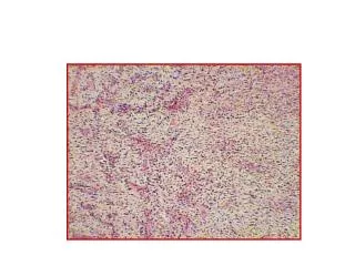
Biopsy techniques • Get normal tissue with abnormal tissue • If surface lesion--don’t need to go too deep • If swelling or mass--the deeper the better • Don’t biopsy the middle of an ulcer • Lasso technique • Mark with sutures • Punch biopsy--5mm minimum • Include picture and differential with as much clinical info as possible--this is very important • Contact the pt right away with results!!
Traumatic (simple) bone cyst • Not a true cyst; posterior mandible; asymptomatic or painless swelling • X-ray: well-defined unilocular radiolucency; “scalloped” appearance in multiple teeth involvement • Histo: fibrovascular CT & trabecular bone; cyst may be empty • Tx: surgical exploration & tissue submission; good prognosis, rapid new bone formation • DD: periapical granuloma, periapical cyst, periapical cemento-osseous dysplasia, periapical scar, dentin dysplasia type 1 (page 804)
Granular Cell Tumor-no diff. diagnosis in book-nodular mass under skin or mucosa-tongue and buccal muscoa-schwann cell origin or neuroendocrine cellstx: local excision
Allergic Stomatitis –Dentifrice Stomatitis-pseudomembranous candidiasis, morsicatio, sloughing traumatic lesion, mouthwash, chemical burn-burning, slight redness to brilliant erythematous lesion, edema possible, superficial aphthous ulcerations possible, stinging tingling, *superficial epithelial sloughing-located at site of contact-dentifrice, medications, lip stick, metals-tx: remove allergen, antihistamines if necessary
Angioedema-no diff. Dx listed in book-diffuse edematous swelling of soft tissue, nontender, solitary or multiple-face, lips, tongue, pharynx, larynx, hands, arms, legs, genitals, buttocks-cause: mast cell degranulation which leads to histamine release and typical IgE hypersensitivity reaction from drugs, foods, plants, dust, heat cold, stress, complement cascade is common in hereditary andioedema-tx: oral antihistamines, intramuscular epi,