Download
1 / 34
410 likes | 1.11k Vues
PRINCIPLES OF MECHANICAL VENTILATION and BLOOD GAS INTERPRETATION. Definitions. Tidal Volume (TV): volume of each breath. Rate: breaths per minute. Minute Ventilation (MV): total ventilation per minute. MV = TV x Rate. Flow: volume of gas per time.

E N D
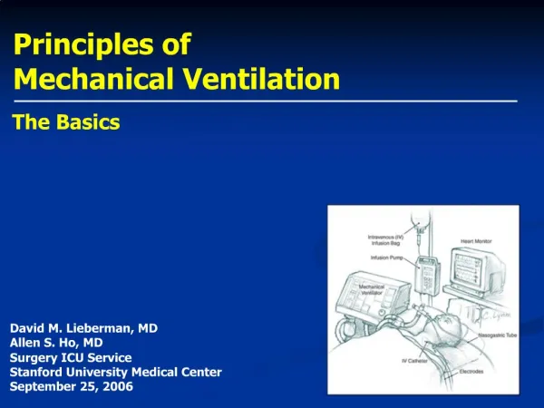
PRINCIPLES OF MECHANICAL VENTILATION and BLOOD GAS INTERPRETATION
Definitions • Tidal Volume (TV): volume of each breath. • Rate: breaths per minute. • Minute Ventilation (MV): total ventilation per minute. MV = TV x Rate. • Flow: volume of gas per time. • Compliance: the distensibility of a system. The higher the compliance, the easier it is to inflate the lungs. • Resistance: impediment to airflow. • SIMV: patient breathes spontaneously between ventilator breaths. Allows patient-ventilator synchrony, making for a more comfortable experience.
Definitions • PIP: maximum pressure measured by the ventilator during inspiration. • PEEP: pressure present in the airways at the end of expiration. • CPAP: amount of pressure applied to the airway during all phases of the respiratory cycle. • PS: amount of pressure applied to the airway during spontaneous inspiration by the patient. • I-time: amount of time delegated to inspiration.
Types of Ventilation • Volume Control • Pressure Control • Pressure Support-CPAP • Pressure-Regulated Volume Control
Volume Control • The patient is given a specific volume of air during inspiration. • The ventilator uses a set flow for a set period of time to deliver the volume: TV (cc) = Flow (cc/sec) x i-time (sec) • The PIP observed is a product of the lung compliance, airway resistance and flow rate. The ventilator does not react to the PIP unless the alarm limits are violated. • The PIP tends to be higher than during pressure control ventilation to deliver the same volume of air. • With SIMV, the patient can breath spontaneously between vent breaths. This mode is often combined with PS.
Pressure Control • Patient receives a breath at a fixed airway pressure. • The ventilator adjusts the flow to maintain the pressure. • Flow decreases throughout the inspiratory cycle. • The pressure is constant throughout inspiration. • Volume delivered depends upon the inspiratory pressure, I-time, pulmonary compliance and airway resistance. • The delivered volume can vary from breath-to-breath depending upon the above factors. MV not assured. • Good mode to use if patient has large air leak, because the ventilator will increase the flow to compensate it.
Changes in ARDS Volume Control Pressure Control
CPAP-Pressure Support • No mandatory breaths • Patient sets the rate, I-time, and respiratory effort. • CPAP performs the same function as PEEP, except that it is constant throughout the inspiratory and expiratory cycle. • Pressure Support (PS) helps to overcome airway resistance and inadequate pulmonary effort and is added on top of the CPAP during inspiration. • The ventilator increases the flow during inspiration to reach the target pressure and make it easier for the patient to take a breath.
Pressure-Regulated Volume Control • In this mode, a target minute ventilation is set. • The ventilator will adjust the flow to deliver the volume without exceeding a target inspiratory pressure. • Decelerating flow pattern. • No change in minute ventilation if pulmonary conditions change. • Can ventilate at a lower PIP than in regular volume control. • Hard to use on a spontaneously breathing patient or one with a large air leak. • Not a “weaning” mode.
Initial Ventilator Settings • Rate: 20-24 for infants and preschoolers 16-20 for grade school kids 12-16 for adolescents • TV: 10-15ml/kg • PEEP: 3-5cm H2O • FiO2: 100% • I-time: 0.7 sec for higher rates, 1sec for lower rates • PIP (for pressure control): about 24cm H2O. • Pressure Support: 5-10cm H2O.
Adjusting The Ventilator • pCO2 too high • pCO2 too low • pO2 too high • pO2 too low • PIP too high
pCO2 Too High • Patient’s minute ventilation is too low. • Increase rate or TV or both. • If using PC ventilation, increase PIP. • If PIP too high, increase the rate instead. • If air-trapping is occurring, decrease the rate and the I-time and increase the TV to allow complete exhalation. • Sometimes, you have to live with the high pCO2, so use THAM or bicarbonate to increase the pH to >7.20.
pCO2 Too Low • Minute ventilation is too high. • Lower either the rate or TV. • Don’t need to lower the TV if the PIP is <20. • PIP <24 is fine unless delivered TV is still >15ml/kg. • TV needs to be 8ml/kg or higher to prevent progressive atelectasis • If patient is spontaneously breathing, consider lowering the pressure support if spontaneous TV >7ml/kg.
pO2 Too High • Decrease the FiO2. • When FiO2 is less than 40%, decrease the PEEP to 3-5 cm H2O. • Wean the PEEP no faster than about 1 every 8-12 hours. • While patient is on ventilator, don’t wean FiO2 to <25% to give the patient a margin of safety in case the ventilator quits.
pO2 Too Low • Increase either the FiO2 or the mean airway pressure (MAP). • Try to avoid FiO2 >70%. • Increasing the PEEP is the most efficient way of increasing the MAP in the PICU. • Can also increase the I-time to increase the MAP (PC). • Can increase the PIP in Pressure Control to increase the MAP, but this generally doesn’t add much at rates <30bpm. • May need to increase the PEEP to over 10, but try to stay <15 if possible.
PIP Too High • Decrease the PIP (PC) or the TV (VC). • Increase the I-time (VC). • Change to another mode of ventilation. Generally, pressure control achieves the same TV at a lower PIP than volume control. • If the high PIP is due to high airway resistance, generally the lung is protected from barotrauma unless air-trapping occurs.
Weaning Priorities • Wean PIP to <35cm H2O • Wean FiO2 to <60% • Wean I-time to <50% • Wean PEEP to <8cm H2O • Wean FiO2 to <40% • Wean PEEP, PIP, I-time, and rate towards extubation settings. • Can consider changing to volume control ventilation when PIP <35cm H2O.
Pulmonary Barotrauma Ventilator-induced lung injury Nosocomial pneumonia Tracheal stenosis Tracheomalacia Pneumothorax Cardiac Myocardial ischemia Reduced cardiac output Gastrointestinal Ileus Hemorrhage Pneumoperiteneum Renal Fluid retention Nutritional Malnutrition Overfeeding Complications
Acute Deterioration • DIFFERENTIAL DIAGNOSES • Pneumothorax • Right mainstem intubation • Pneumonia • Pulmonary edema • Loss of airway • Airway occlusion • Ventilator malfunction • Mucus plugging • Air leak
Tracheal shift Pneumothorax Wheezing Bronchospasm Mucus plugging Pulmonary edema Pulmonary thromboembolism Asymmetric breath sounds Pneumothorax Mainstem intubation Mucus plugging with atelectasis Decreased breath sounds bilaterally Tube occlusion Ventilator malfunction Loss of airway Physical Exam
Elevated peak and plateau pressures Pneumonia Pulmonary edema Pneumothorax Atelectasis Right mainstem intubation Elevated peak pressure, normal plateau pressure Airflow obstruction Mucus plugging Partial tube occlusion Reduced peak and plateau pressure Cuff leak Ventilator malfunction Large bronchopleural fistula Pressure Patterns
Extubation Criteria • Neurologic • Cardiovascular • Pulmonary
Neurologic • Patient must be able to protect his airway, e.g, have cough, gag, and swallow reflexes. • Level of sedation should be low enough that the patient doesn’t become apneic once the ETT is removed. • No apnea on the ventilator. • Must be strong enough to generate a spontaneous TV of 5-7ml/kg on 5-10 cm H2O PS or have a negative inspiratory force (NIF) of 25cm H2O or higher. • Being able to follow commands is preferred.
Cardiovascular • Patient must be able to increase cardiac output to meet demands of work of breathing. • Patient should have evidence of adequate cardiac output without being on significant inotropic support. • Patient must be hemodynamically stable.
Pulmonary • Patient should have a patent airway. • If no air leak, consider decadron and racemic epinephrine. • Pulmonary compliance and resistance should be near normal. • Patient should have normal blood gas and work-of-breathing on the following settings: • FiO2 <40% • PEEP 3-5cm H2O • Rate: 6bpm for infants, 2bpm for toddlers, CPAP/PS for 1hr for older children and adolescents • PS 5-8cm H2O • Spontaneous TV of 5-7ml/kg
Blood Gas Interpretation NORMAL VALUES Arterial Venous Capillary pH 7.4 (7.38-7.42) 7.36 (7.31-7.41) 7.35-7.40 pO280-100 mm Hg 35-40 mm Hg 45-60 mm Hg pCO235-45 mm Hg 41-52 mm Hg 40-45 mm Hg Sat >95% on RA 60-80% on RA >70% HCO322-26 mEq/L 22-26mEq/L 22-26mEq/L BE -2 to +2 -2 to +2 -2 to +2
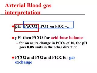
Rules Of Interpretation • ∆ in pCO2 of 10mm Hg should ∆ pH by 0.08. • pH ∆ of 0.15 is equal to ∆ in HCO3 of 10mEq/L. • Normal pCO2 in the face of respiratory distress is a sign of impending respiratory failure.
Acid-Base Diagram From Goldberg, M., Green, S.B., Moss, M.L., et al.: JAMA 223:269-275, 1973
Respiratory Disturbances • Acute respiratory acidosis occurs when CO2 is retained acutely. • Chronic respiratory acidosis occurs when the retained CO2 gets buffered by renal retention of HCO3. The pH is higher than in acute respiratory acidosis, but it is still <7.4. • Acute respiratory alkalosis occurs when CO2 is blown off acutely. • Chronic respiratory alkalosis occurs when the reduction of CO2 is compensated for by the renal excretion of HCO3. The pH is lower than in acute respiratory alkalosis, but it is still >7.4.
Metabolic Disturbances • Acute metabolic acidosis gets compensated by CO2 reduction within 12-24 hours. The pH is still usually <7.4. • Metabolic alkalosis is rare. Usual causes are pyloric stenosis, chronic diuretic use, and bicarbonate infusions. • Otherwise healthy people do not usually retain CO2 to compensate for metabolic alkalosis. • Patients who are severely dehydrated or have lung disease will retain CO2 to compensate for metabolic alkalosis.
Hypoxemia There are five reasons for hypoxemia: • FiO2 too low (high altitude) • Global alveolar hypoventilation • Right-to-left shunts • V/Q mismatch • Incomplete diffusion