Understanding Gene Mutations and the Genetic Code
This overview covers essential concepts related to gene mutations and the genetic code, focusing on the structure of prokaryotic genes, including promoters and ribosome binding sites. It explains how DNA generates RNA for protein synthesis and highlights the genetic code's properties, such as triplet codons, degeneracy, and stop signals. Additionally, it discusses mutation types, including spontaneous and environmentally induced mutations, and the mutation rate in human DNA. This comprehensive approach provides a foundation for understanding genetic variations and their implications.

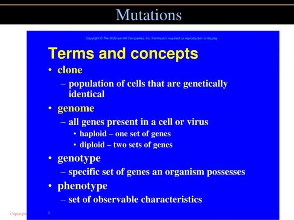
Understanding Gene Mutations and the Genetic Code
E N D
Presentation Transcript
What is a gene? Prokaryotic Genes PROMOTER 3’ 5’ antisense ---TTGACAT------TATAAT-------AT-/-AGGAGGT-/-ATGCCC CTT TTG TGA ---AACTGTA------ATATTA-------TA-/-TCCTCCA-/-TAC GGG GAA AAC ATT sense (-35) 3’ (-10) 5’ RIBOSOME BINDING SITE 3’ 5’ U-/-AGGAGGU-/-AUGCCC CUU UUG UGA Met Pro leu leu stp When ALL OF THESE RULES ARE SATISFIED THEN THEN A PIECE OF DNA WILL GENERATE A RNA WHICH WILL BE READ AND TRANSLATED INTO A PROTEIN.
Reading the genetic code U U U A A A U A C Lys Phe Met 5’ 3’ A T G T T T A A A T A G C C C C A T A A A T T T C T A G G G 3’ 5’ 5’ 3’ A T G T T T A A A T A G C C C 5’ 3’ 5’ 3’ A U G U U U A A A U A G C C C C A T A A A T T T C T A G G G 3’ 5’ A U G U U U A A A U A G C C C S T P
No Gaps 5’ 5’ 3’ 3’ A U G A U G U U U U U U A A A A A A U A G U A G C C C C C C A A U U U A U U U A A A U A C U A C Asn Met Lys Phe Met Leu S T P
No overlaps 5’ 5’ 3’ 3’ A U G A U G A A A A A A C C C C C C U A G U A G C C C C C C U U U U G G G G G U U U U A C U A C Lys Met Pro Lys Met Trp S T P
The GENETIC CODE The code is a three letter code. Second letter U C A G UUU UUC UUA UUG CUU CUC CUA CUG AUU AUC AUA AUG GUU GUC GUA GUG UCU UCC UCA UCG CCU CCC CCA CCG ACU ACC ACA ACG GCU GCC GCA GCG UAU UAC UAA UAG CAU CAC CAA CAG AAU AAC AAA AAG GAU GAC GAA GAG UGU UGC UGA UGG CGU CGC CGA CGG AGU AGC AGA AGG GGU GGC GGA GGG U C A G U C A G U C A G U C A G Phe Tyr Cys U Ser STOP STOP Leu Trp His C Arg Pro Leu Gln Third letter First letter Asn Ser Ile A Thr Arg Lys Met Asp G Val Ala Gly Glu
The code Ser Ser Ser Ser Ser AGG AGU UCG AGG AGG 3 amino acids are specified by 6 different codons 5 amino acids are specified by 4 different codons 1 amino acid is specified by 3 different codons 9 amino acids are specified by 2 different codons 2 amino acids are specified by 1 different codons The degeneracy arises because More than one tRNA specifies a given amino acid A single tRNA can base-pair with more than one codon tRNAs do not normally pair with STOP codons ----UCC------UCA------AGC ----UCC------UCA------
The Genetic Code Properties of the Genetic code: 1- The code is written in a linear form using the nucleotides that comprise the mRNA 2- The code is a triplet: THREE nucleotides specify ONE amino acid 3- The code is degenerate: more than one triplet specifies a given amino acid 4- The code is unambiguous: each triplet specifies only ONE amino acid 5- The code contains stop signs- There are three different stops 6- The code is comma less 7- The code is non-overlapping
Generation of mutations Spontaneous mutations Replication induced mutations of DNA Usually base substitutions (Most errors are corrected) Meiotic crossing over can induce mutations Small additions and deletions AND Large changes as well Environment induced changes Exposure to physical mutagens - Radioactivity or chemicals Depurination (removal of A or G) Repair results in random substitution during replication Deamination (removal of amino group of base) (nitrous acid) Cytosine--uracil--bp adenine--replication-- Oxidation (oxoG) guanine--oxoguanine--bp adenine--replication -- Base analog incorporation during replication BU-T Intercalating agents
Methods used to study mutations Gross chromosomal changes- deletions, insertions, inversions, translocations Cytology- microscopy- karyotype Small mutations Small deletions, insertions and point mutations Recombinant DNA technologies
Mutation rate There are approximately 1013 cells in the human body Each cell receives 10,000 DNA lesions per day (Lindahl and Barnes 2000). Most pervasive agent is UV. 100,000 lesions per exposed cell per hour (Jackson and Bartek 2009). Ionizing agents (X-rays/g-rays) are most toxic because they generate double strand breaks (Ward 1988). Chromosome instability (gain or loss of entire segments) is frequent - 40% of imbalances are entire arm imbalance while 45% are terminal segment imbalance (double strand break, nondysjunction etc) Sequencing 179 humans as part of the 1000 genome project: On average, each person is found to carry approximately 250 to 300 loss-of-function variants in genes of which 50 are in genes previously implicated in inherited disorders. 1.3 million short indels (1-10,000 bp) were identified and 20,000 large (>10,000 bp) variants were identified. Variation detected by the project is not evenly distributed across the genome: certain regions, containing repetitive sequences (sub-telomeres etc), show high rates of indels.
Sequencing the whole genomes of a family (2010 Science 328 636). 98 crossovers in maternal genome 57 crossovers in paternal genome Mutation rate is 1x10⁻⁸ per position per haploid genome (human genome is 3x10⁹ bp) It was calculated that there are ~70 new mutations in each diploid human genome Some sites such as CpG sites mutate at a rate 11 times higher than other sites Exome sequencing of 2440 individuals (Science 2012 337 40) Each person has ~100 loss of function mutations (~35 nonsense). 20 loss of function mutations are homozygous Some alterations in sequence concentrate in specific geographic populations Rare changes are population specific and their frequencies vary for each geographic population
Chromosomes and chromosome rearrangements Cytogenetics is the study of genetics by visualizing chromosomes. This area of research is germane to several areas of biological research. Cytogenetics has been fundamental to understanding the evolutionary history of a species (for example, although the Chimp and the human are morphologically very different, at the level of the chromosome (and DNA sequence) they are extremely similar. H = human C= chimp G = Gorilla O = Orang utang
Karyotype Chromosomes are classified by size, centromere position and banding pattern: Shown below is the human karyotype (description of the chromosome content of a given species) Karyotype is the chromosome description of length, number, morphology. Karyotype analysis is extremely important in medicine. Alternations in karyotypes are linked to birth defects and many human cancers. Metacentric- centromere in the middle Acrocentric- centromere off center telocentric centromere at one end
Banding patterns Specialized stains produce unique banding patterns along each chromosome. Banding patterns are extremely useful for detecting abnormalities in chromosome structure. For many of the chromosome stains- the molecular basis of the banding patterns is unclear. Nonetheless these techniques remain fundamental in many areas of genetic research
MU to bp Genetic maps are based on recombination frequencies and describe the relative order and relative distance between linked genes. Remember genes reside on chromosomes. So what we would like to know is where are the genes located on the chromosomes 22% Rf = 22MU What does this mean in terms of chromosomes and DNA?
Physical maps Physical maps provide information concerning the location of genes on chromosomes Where are the genes on chromosomes? Cytological studies have been successfully used to map genes to specific regions of a chromosome. For example in Drosophila in some cells the chromosomes become highly replicated and exhibit very characteristic banding patterns:
In situ hybridization Salivary glands Squash on slide Denature/Stain polytene chromosomes label gene probe (you can only use this method if you have the gene cloned) Hybridize probe to polytene chromosomes Autoradiography
Chromosome loss Chromosome instability- Elevated gain or loss of complete chromosomes Frequent in tumors Frequent in in vitro fertilized embryos Gross chromosomal rearrangements during in vitro fertilization. 40% of embryos carried entire chromosome imbalance Gain or loss of segments of chromosomes CNV- copy number variation of chromosome segments 5% of individuals genome displays CNV Synuclein gene CNV is involved in Parkinsons Many cancers- malignant cells most often gain additional copies of chromosome segments- genes in these segments are mis-expressed or mis-express other genes) Microarray hybridization of DNA from tissues of identical twins- differences at several loci seen (Bruder et al 2008) Microarray hybridization of DNA from different tissues of single individual (Piotrowski et al., 2008) Gross chromosomal rearrangements during in vitro fertilization 55% of embryos carried terminal imbalance (sub-telomere loss) (Vanneste et al., 2009) -microarray based screen of IVF 35 embryo
Gross chromosomal changes The Cri du chat syndrome in humans is a result of a deletion in the short arm of chromosome 5. This was determined by comparing banding patterns with normal and Cri du Chat individuals Types of chromosome rearrangements that can be studied by karyotype analysis: GROSS CHROMOSOMAL CHANGES Deletions, Duplications, Inversions, Translocations
DDIT A____B____C________D____E____F A____B____C________D____F A____B____C________D____E____E____F A____B____C________E____D____F A____B____C________D____E____F A____B____C________D____L H____I____J________K____L H____I____J________K____E____F Normal Chromosome Deletions (deficiency) Duplications Inversions Translocation
Insertion and deletions are frequent: Sequencing 179 humans as part of the 1000 genome project: On average, each person is found to carry approximately 250 to 300 loss-of-function variants in genes of which 50 are in genes previously implicated in inherited disorders. 20,000 large structural variants were identified and 1.3 million short indels were identified. Variation detected by the project is not evenly distributed across the genome: certain regions, such as subtelomeric regions, show high rates of variation. 1 in 500 children have reciprocal translocation but Such translocations are usually harmless. (However gametes produced by the children will have defects). 1 in 50 children have inversions (small and large). The heterozygous inversion carrier generally show no adverse phenotype (but produce abnormal meiotic products from crossing-over in the inversion loop).
Deletions Deletions are often detected cytologically by comparing banding patterns between the normal and the partially deleted chromosomes Deleted segment Chromosome no female deletion chromosome1 Band 46,XX, del(1)(q24q31) Female with a deletion of chromosome 1 on the long arm (q) between bands q24 to q31.
In many instances deletions are too small to be detected cytologically. In these instances genetic/molecular techniques are used. Since cytological deletions remove a contiguous set of genes, there is a high probability that an essential gene will be deleted. Therefore deletions will survive as heterozygotes and not homozygotes. A____B________C____D Normal A____B________C____D A____________C____D Homologous deletion (Lethal?) A____________C____D A____________C____D Heterologous deletion (NOT Lethal) A____B________C____D
Consequences of deletions A+_____B+_____C+___________D+ Normal A+_____B+_____C+___________D+ In individuals heterozygous for the deletion, pairing is disrupted in the regions surrounding the deletion. Therefore recombination is also significantly reduced in these regions. B+ A+____/ \_____C+___________D+ A+___________C+___________D+ Genotype A+_____b______c____________D+ Normal A+_____B+_____C+___________D+ A deletion on one homologue unmasks recessive alleles on the other homologue. The effect is called pseudo-dominance. A+____b______c____________D+ A+___ _____C+___________D+
Deletions in X Females in Drosophila XX Males in Drosophila XY or XO Deletion series phenotype sick dead sick
Changes in chromosome structure Deletions: Hemizygosity from large deletions results in lethality- even the smallest cytologically defined deletions take out tens of 1,000's of bps and are likely to remove essential genes. 2. Organisms can tolerate hemizygosity from small but not large deletions. The reason for this is not entirely clear and is placed under the rubric of disrupting the overall ratio of gene products produced by the organism
Duplications A____B____C________D____E____F A____B____C________D____E____E____F normal Duplication Individuals bearing a duplication possess three copies of the genes present in the duplicated region. In general, for a given chromosomal region, organisms tolerate duplications much better than deletions. 46,XY, dup(7)(q11.2q22) Male with a duplication of chromosome 7 on the long arm (q) between bands 11.2 to 22
Tandem duplications- Important class of duplications!!! This is a case in which the duplicated segment lies adjacent to the original chromosomal segment A B C D ------ A B CB CB CB C D Once a tandem duplication arises in a population, even more copies may arise because of asymmetrical pairing at meiosis. Remember when the homologs pair during prophase of meiosis I, they line up base-pair for base pair. Duplications lead to mistakes in this pairing mechanism
Proper pairing: A____B____C____B____C____D____E A____B____C____B____C____D____E A____B____C____B____C____D____E A____B____C____B____C____D____E Inappropriate pairing: A____B____C____B____C____D____E A____B____C____B____C____D____E A____B____C____B____C__-----------__D____E A____B____C____B____C__-----------__D____E
Tandem duplications expand by mistakes in meiosisI during pairing a b c b c d e B C E B A C D
Tandem duplications expand by mistakes in meiosis during pairing Paired non-sister chromatids B C E A B C D c b a d e c b 33
What happens if you get a crossover after mis-pairing in meiosisI? A B C B C D A B C B C D c b b c c b a b d a c d B C E A B C D A B C B C D A B C B C B C D c b a d e A B C D c b A B C B C D
The four meiotic products of a crossover between regions B and C: A-B-C-B-C-D-E A-B-C-D-E A-B-C-B-C-B-C-D-E A-B-C-B-C-D-E This process may repeat itself many times, such that a small fragment of the genome is repeated 10,000 times.
An example of this is near the centromeres of the Drosophila genome: If you look at the DNA sequence in this region it consists of small 5-10 bp sequences (AATAC)n repeated 1,000s of times. It is believed to have arisen from unequal crossing over. Repetitive DNA- cell does not like it- They try to reduce recombination of repetitive DNA by packaging the DNA with proteins to form heterochromatin- cold spots of recombination along the chromosome
Duplications provide additional genetic material capable of evolving new function. For example in the above situation if the duplication for the B and C genes becomes fixed in the population- the additional copies of B and C are free to evolve new or modified functions. This is one explanation for the origin of the tandemly repeated globin genes in humans. Each of these has a unique developmental expression pattern and provides a specialized function. The hemoglobin in fetus has a higher affinity for oxygen since it acquires its oxygen from maternal hemoglobin via competition
Inversion Chromosomes in which two breaks occur and the resulting fragment is rotated 180 degrees and reinserted into the chromosome. Inversions involve no change in the amount of genetic material and therefore they are often genetically viable and show no abnormalities at the phenotypic level. Gene fusions may occur Inversions are defined as to whether they span the centromere Paracentric inversions do not span the centromere: A B C D E A B DC E Pericentric inversions span the centromere: A CB D E In a pericentric inversion one break is in the short arm and one in the long arm. Therefore an example might read 46,XY,inv(3)(p23q27). A paracenteric inversion does not include the centromere and an example might be 46,XY,inv(1)(p12p31).
Homologs which are heterozygous for an inversion have difficulties pairing in meiosis. During pairing homologous regions associate with one another. Consequently individuals heterozygous for an inversion will form a structure known as an inversion loop. Crossover within inverted region? A---B---C---D---E---F---G A’--B’---C’---D’--E’---F’---G’ A---B---C---D---E---F---G A’--B’---C’---E’---D’--F’---G’ D E D’ E’ A B C F G ‘F G’ A’ B’ C’
The consequence of crossover within a paracentric inversion a-b-c d-e f-g a-b-c d-e f-g a-b-c e-d f-g During meiosis, pairing leads to formation of an inversion loop This is a problem if crossing over occurs within the inversion D E D’ E’ A B C F G ‘F G’ A’ B’ C’ A-B-0-C-D-E’-C’--0--B’-A’ dicentric-fragmentation G-F-E-D’-F’-G’ acentric- no segregation
The consequence of crossover within a pericentric inversion (one that spans the centromere). a-b-c d-e f-g a-b-c d-e f-g a-b-c e-d f-g During meiosis, pairing leads to formation of an inversion loop This is a problem if crossing over occurs within the inversion D E D’ E’ A B C F G ‘F G’ A’ B’ C’ A-B-C-D-0-E’-C’-B’-A’ fragment G-F-E-0-D’-F’-G’ fragment
Paracentric inversion crosses over with a normal chromosome, the resulting chromosomes are an acentric, with no centromeres, and a dicentric, with 2 centromeres. • The acentric chromosome isn't attached to the spindle, so it gets lost during cell division, and the dicentric is usually pulled apart (broken) by the spindle pulling the two centromeres in opposite directions. These conditions are lethal. • Pericentric inversion crosses over with a normal chromosome, the resulting chromosomes are duplicated for some genes and deleted for other genes. (They do have 1 centromere apiece though). • The gametes resulting from these do not produce viable progeny. • Thus, either kind of inversion has lethal results when it crosses over with a normal chromosome. • The only offspring that survive are those that didn't have a crossover or crossed over in regions outside the inversion. • Thus when you count the offspring you only see the non-crossovers, so it appears that crossing over has been suppressed.
What are the consequences of crossing-over in an individual homozygous for an inversion? Genotype for normal individual A B 0 C D E F G A B 0 C D E F G Genotype of an individual heterozygous for an inversion: A B 0 C D E F G A B 0 C F E D G Genotype of an individual homozygous for an inversion: A B 0 C F E D G A B 0 C F E D G
Translocations A segment from one chromosome is exchanged with a segment from another chromosome. Chromosome 1 A B C D E F ----------------------0----------------------- ----------------------0----------------------- A B C D E F Chromosome 2 O P Q R S T ----------------------0----------------------- ----------------------0----------------------- O P Q R S T Reciprocal translocation A B C D S T ----------------------0----------------------- O P Q R E F ----------------------0----------------------- This is more specifically called a reciprocal translocation and like inversions (and unlike duplications and deficiencies) no genetic material is gained or lost in a reciprocal translocation. Non-reciprocal translocations may also occur
long arms of chromosome 7 and 21 have broken off and switched places. So you can see a normal 7 and 21, and a translocated 7 and 21. This individual has all the material needed, just switched around (translocated), so they should have no health problems. However there can be a problem when this person has children. Remember that when the gametes are made, each parent gives one of each chromosome pair. What would happen if this person gave the normal seven and the 21p with 7q attached? There are three copies of 7q instead of two. And there is only one copy of 21q t(11;18)(q21;q21) translocation between chromosomes 11 and 18 at bands q21 and q21 Philadelphia chromosome: t(9;22)(q34;q11).
As with inversions, individuals heterozygous for a reciprocal translocation will exhibit abnormalities in chromosome pairing A B C D E F ----------------------0----------------------- ----------------------0----------------------- A B C D S T O P Q R S T ----------------------0----------------------- ----------------------0----------------------- O P Q R E F Notice this individual has the normal amount of genetic material (two copies of each gene). However it is rearranged. If the translocated fragment contains a centromere, you could get dicentri and acentric chromosomes How will translocated chromosomes pair in meiosis?
F F N1 T1 E E A B C D R Q P O A B C D R Q P O S S T2 N2 T T Homologous regions associate with one another. These chromosomes will follow Mendel's rule of independent of assortment. In this instance one must focus on the centromere There are three possible patterns of segregation. Normal Pairing of 10 chromosomes in maize Chr8-9 translocation
F F N1 T1 • Alternate segregation: • キN1 and N2 segregate to one pole • キT1 and T2 segregate the other pole • These gametes have the normal haploid gene content: one copy of each gene and are normal • Adjacent segregation: • キN1 and T1 segregate to one pole • キT2 and N2 segregate to the other pole • These gametes are anueploid: they are missing some genes and duplicated for other genes. • Adjacent segregation • キN1 and T2 segregate to one pole • キN2 and T1 segregate to one pole • Therefore, in a translocation heterozygote, some of the gametes are viable and some are inviable. E E A B C D R Q P O A B C D R Q P O S S T2 N2 T T
F F N1 T1 E E A B C D R Q P O A B C D R Q P O Reciprocal translocations result in genes that are known to map to different chromosomes but behave as linked genes. Under normal circumstances genes E and R assort independently because they are on different chromosomes. However in a translocation they will behave as closely linked genes and segregate together. S S T2 N2 T T
Translocations (and inversion) breakpoints sometimes disrupt an essential gene. That is the break occurs in the middle of a gene. In fact because of this, a number of specific translocations are causally associated with specific human cancers. The inherited disease Duchenne muscular dystrophy was mapped through a translocation that specifically disrupted this gene.