Download
1 / 55
580 likes | 807 Vues
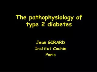
The pathophysiology of type 2 diabetes. Jean GIRARD Institut Cochin Paris. Genetic factors. Insulin-resistance. Hyperinsulinemia. Acquired factors. Compensation. Normal glucose tolerance. Acquired factors. Genetic factors. Gluco-lipotoxicity. ß-cell deficiency.

E N D
The pathophysiology of type 2 diabetes Jean GIRARD Institut Cochin Paris
Genetic factors Insulin-resistance Hyperinsulinemia Acquired factors Compensation Normal glucose tolerance Acquired factors Genetic factors Gluco-lipotoxicity ß-cell deficiency Insulin-resistance Glucose production Insulin secretion Type 2 diabetes
Causes of hyperglycemia in type 2 diabetes Liver Glucose Muscle Insulin Glucagon Pancreas
The euglycemic clamp Plasma insulin (mU/ml) 100 50 0 Exogenous glucose (mg/min/kg) 10 5 0 Plasma glucose (md/dl) 100 90 80 70 60 0 1 2 3 hours
Insulin resistance in type 2 diabetes Hepatic glucose production Peripheral glucose utilisation Control T2D T2D Control 200 0 100 0 100 50 Plasma insulin (µU/ml)
Insulin-stimulated glucose uptake and glycogen synthesis are reduced in Type 2 diabetes Glucose uptake (mg/min/kg) 3 2 Glycogen synthesis Glycolysis 1 Oxidation 0 Control Type 2 diabetes
Non-oxidative glucose metabolisme in skeletal muscle Glucose Glucose transport Glucose Hexokinase II Glucose-6-P Glycolysis Glycogen synthase Pyruvate Glycogen
Effect of insulin on glucose transport in skeletal muscle of type 2 diabetes 3-O-methylglucose transport (mmol/h/ml cell water) 2.5 Control 2.0 Type 2 diabetes 1.5 1.0 0.5 0 0 100 200 400 1000 Insulin (mU/ml)
Glycogen synthesis in skeletal muscles during a hyperglycemic hyperinsulinemic clamp Glycogen concentration in gastrocnemius (mmol/kg) 20 Control 15 10 Type 2 diabetes 5 0 0 20 40 60 80 100 120 Minutes
Conclusions Skeletal muscles are responsible for the decreased whole body insulin-stimulated glucose uptake Glucose transport is the rate-limiting step of insulin-stimulated glucose metabolism in skeletal muscle Insulin-stimulated skeletal muscle glycogen metabolism is reduced in type 2 diabetes
Autophosphorylation Tyrosine kinase activity Binding a a a a a Insulin a Extracellular b b b b b b b P-Tyr Tyr-P P-Tyr Tyr Tyr Tyr-P Intracellular ATP IRS-Tyr-P IRS Metabolic effects
Insuline PI 3 kinase p85 p110 Tyr-P Tyr-P P-Tyr Tyr-P IRS-1 PDK-1 Tyr-P Tyr-P Tyr-P Protéine kinase C Protéine kinase B Metabolic effects Glucose transport Glycogen synthesis Inhibition of glucose production
IRS-1 tyrosine phosphorylation in human skeletal muscle % of basal values 8 Basal Clamp 4 0 Control Type 2 diabetes
IRS-1 associated PI 3 kinase in human skeletal muscle % of basal values 500 Basal Clamp 250 0 Control Type 2 diabetes
Defects in insulin-signaling pathways in Type 2 diabetes The insulin receptor number is reduced by 20%, but this is compensated by hyperinsulinemia The tyrosine phosphorylation of IRS-1 and the activation of PI 3 kinase are decreased in Type 2 diabetes Increased activity of tyrosine phosphatases ? Serine phosphorylation of IRSs ?
Factors responsible for the decrease in insulin signaling in Type 2 diabetes Defect Factors responsible Insulin receptor number Increased plasma insulin Tyrosine kinase activity Serine Phosphorylation of IRS Glucose transport Hyperglycemia, decreased Glut4 translocation
Insulin Hyperglycemia Tyr-P Tyr-P IRS-1 Ser-P Protein Kinase C Tyr-P Tyr-P Tyr-P Decreased association with PI 3 Kinase Glucose transport
The hexosamine pathway Glucose Glucose-6-P Glutamine:fructose-6-P amidotransferase Fructose-6-P Glucosamine-6-P Glutamine Glutamate Glycolysis N-acetyl-glucosamine-6-P Pyruvate UDP-N-acetyl-glucosamine
Possible role of metabolites of the hexosamine pathway in insulin resistance due to chronic hyperglycemia Glucose Glucose G-6-P G-1-P Glycogène GFA Glucosamine-6-P F-6-P N-acétyl-Glucosamine-6-P Pyruvate UDP-N-Acétyl-glucosamine Glycoprotéines GFA = Glutamine:fructose-6-P amidotransferase
The O-GlcNac glycosylation of glycogen synthase results in reduced activation in response to insulin Glycogen synthase activity % of total 100 Insulin 50 Insulin + glucosamine 0 10 100 1000 G-6-P (mM)
Insulin resistance in type 2 diabetes Adipose tissue lipolysis 100 T2D Control 0 30 0 100 Plasma insulin (µU/ml)
Type 2 diabetics have high plasma FFA all along the day Plasma FFA (mmol/l) 800 Type 2 diabetes 600 400 200 Control 0 8 10 12 14 16 18 20 Hours
Fatty acid-induced insulin resistance : Randle’s hypothesis 1963 FFA Glucose Glucose Fatty acyl-CoA HK G-6-P Acetyl-CoA NADH Citrate PFK PDH Mitochondria Pyruvate
Potential steps controlling muscle glucose metabolism in response to FFA Glucose transport Hexokinase Glycogen synthase Glucose Glucose Glycogen G-6-P Metabolite levels during the clamp 100 Arbitrary units 50 0 Control FFA Control FFA Control FFA
Fatty acid-induced insulin resistance : Shulman 1999 FFA Insulin Fatty acyl-CoA IRS-SerP Protein kinase Cf PI 3 Kinase Glucose transport
Adipose tissue of type 2 diabetics Résistine Visfatine Adiponectine TNFa IL-6 Insulin resistance Insulin sensitivity
Insulin TNFa Sphingomyelinase Tyr-P Ceramides IRS-1 Protein Kinase C PTPase Tyr-P Ser-P Tyr-P Tyr-P Tyr-P Decreased association with PI 3 Kinase Biological effects
Cytokine-induced insulinoresistance Insulin TNF, IL-6 IRS- Ser P JNK IKKß PI 3 Kinase SOCS JNK = Jun kinase Glucose transport Metabolic effects IKKß = Inhibitor of kappa B kinase ß SOCS =Suppressor of cytokine signaling
L’insulinorésistance induite par les cytokines Salicilate TNF, IL-6 Insuline IRS- Ser P IKKß PI 3 Kinase Effets métaboliques de l’insuline IKKß = Inhibitor of kappa B kinase ß
Postabsorptive glucose concentration is related to hepatic glucose production in type 2 diabetes Hepatic glucose production (mg/min/h) 4 3 2 1 0 0 100 200 300 400 Postabsorptive glucose concentration (mg/min/h)
Gluconeogenesis is responsible for increased hepatic Glucose production in type 2 diabetes Hepatic glucose production (mg/min/kg) 4 3 Glycogenolysis 2 1 Gluconeogenesis 0 Control Type 2 diabetes
Factors responsible for increased hepatic glucose production in Type 2 diabetes 1- Liver insulin resistance 2- Increased plasma glucagon levels 3- Increased plasma FFA levels
Insulin resistance in type 2 diabetes Hepatic glucose production (mg/min/kg) 4 2 T2D Control 0 50 0 100 Plasma insulin (µU/ml)
Type 2 diabetics have high plasma glucagon despite hyperglycemia Plasma glucagon (pg/ml) Type 2 diabetes 200 150 100 Control 50 0 8 10 12 14 16 18 20 Hours
Glucagon in type 2 diabetes Increased mass of A cells • Increased ratio A cells/B cells • • Hyperglucagonemia despite hyperglycemia • Increased secretion in response to amino-acids • Secretion of glucagon is less inhibited in response to glucose The impairement of glucagon secretion precedes the appearance of type 2 diabetes •
Factors responsible for hyperglucagonemia in Type 2 diabetes Insulin resistance of A cells ? • No: The impairement of A cells is not corrected by appropriate insulin-therapy • Chronic hyperglycemia desensitizes A cell ? Possible : glucagon secretion is corrected by normalization of glycemia in response to phlorizine • Mechanisms responsible for glucose « blindness » of A cells ?
Consequences of chronic hyperglucagonemia on hepatic glucose production in type 2 diabetes Increased transcription of genes coding for gluconeogenic enzymes : for exemple PEPCK Glucose production mainly due to gluconeogenesis Gluconeogenesis is less sensitive than glycogenolysis to the inhibition by insulin : Insulin resistance The absence of inhibition of glucagon secretion in the postprandial state induced glucose intolerance due to the non-suppression of hepatic glucose production
Glycogenolysis is very sensitive whereas gluconeogenesis is insensitive to an increase in portal insulin 6 Glycogenolysis 5 4 Basal Glycogenolysis Gluconeogenesis (mg/min/kg) 3 2 Glucogeogenesis 1 0 0 10 20 30 40 50 Portal insulin (mU/ml)
Exogenous glucose appearance (mg/min/kg) Plasma glucose (mg/dl) 4 Type 2 diabetes 400 3 Type 2 diabetes 300 2 200 1 100 Control Control 0 0 -60 0 60 120 180 240 360 -60 0 60 120 180 240 360 Minutes Minutes Glucose Glucose Glucose intolerance after oral glucose administration In type 2 diabetes
Glucose utilization Hepatic glucose production (mg/min/kg) (µmol/min/kg) 8 4.0 Type 2 diabetes Type 2 diabetes 6 3.0 4 2.0 2 1.0 Control Control 0 0 -60 0 60 120 180 240 360 -60 0 60 120 180 240 360 Minutes Minutes Glucose Glucose Glucose intolerance after oral glucose administration is due to non-suppression of hepatic glucose production
Plasma glucagon (pg/ml) Plasma insulin (µU/ml) 100 200 Control Type 2 diabetes Type 2 diabetes 75 150 50 100 25 50 Control 0 0 -60 0 60 120 180 240 360 0 60 120 180 240 360 -60 Minutes Minutes Glucose Glucose Glucose intolerance after oral glucose administration is due to non-suppression of plasma glucagon and to the absence of early insulin secretion
What could be expected from an inhibition of glucagon secretion in type 2 diabetes ? Plasma glucose Hepatic glucose production (mg/dl) (µmol/min/Kg) Type 2 diabetes 400 4.0 300 Type 2 diabetes 3.0 200 2.0 GLP-1 100 GLP-1 1.0 0 0 -60 0 60 120 180 240 360 -60 0 60 120 180 240 360 Minutes Minutes Glucose GLP-1 Glucose GLP-1
Hepatic glucose production (µmol/min/kg) Glucose Glucose Répaglinide Répaglinide What could be expected from restoring the first phase of insulin secretion, in type 2 diabetes ? Plasma glucose (mg/dl) 400 4.0 Type 2 diabetes Type 2 diabetes 300 3.0 Répaglinide 200 2.0 Répaglinide 100 1.0 0 0 -60 0 60 120 180 240 360 -60 0 60 120 180 240 360 Minutes Minutes
Type 2 diabetics have high plasma FFA Plasma FFA (mmol/l) 800 Type 2 diabetes 600 400 200 Control 0 8 10 12 14 16 18 20 Hours
Visceral fat and insulin resistance 100 Insulin Sensitivity mmol/min/kg) 20 20 50 30 40 % visceral fat
Hepatic fatty acid oxidation provides co-factors essential for gluconeogenesis Pyruvate OAA PEP 3-PGA 1,3-DPG GAP Glucose ATP GTP ATP NADH ATP Acetyl-CoA ATP NADH Fatty acid oxidation
Role of Free Fatty Acids in Hyperglycemia Adipose tissue Muscle Liver FFA Glucose utilization Gluconeogenesis Hyperglycemia
Glucose-induced insulin secretion is decreased in chronically hyperglycemic Type 2 diabetic patients Mean plasma insulin during the OGTT (mU/ml) 100 80 60 40 Fasting plasma insulin (mU/ml) 20 0 80 100 120 140 160 180 Fasting plasma glucose levels (mg/dl)
Compensation of insulin-resistance by pancreatic ß-cells Increased insulin secretion Increased ß-cell mass Replication of pre-existing ß-cells, neogenesis of ß-cells Alteration of proliferation or survival of ß-cells
Pancreatic ß cells from type 2 diabetic patients Functional defect • Pulsatility • First phase • Glucose-induced insulin secretion Decrease in ß-cell mass • Genetic factors (HNF1, HNF4, Kir6.2, TCF7L2, Mitochondrial genes) • Environmental factors (Gluco-lipotoxicity, physical inactivity)