MR Airway Pressure Device
130 likes | 299 Vues
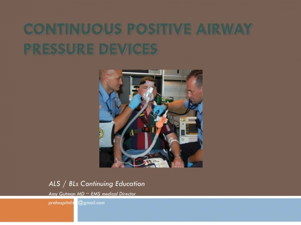
MR Airway Pressure Device. Group Members Laura Sheehan Kevin Johnson Jon Cappel Noelle Simatic Client Dr. Victor Haughton Advisor Prof. Mitch Tyler. Abstract.

MR Airway Pressure Device
E N D
Presentation Transcript
MR Airway Pressure Device Group Members Laura Sheehan Kevin Johnson Jon Cappel Noelle Simatic Client Dr. Victor Haughton Advisor Prof. Mitch Tyler
Abstract An airway pressure device was designed for use in an MR imager. This device will assist our client in studying CSF flow during Valsalva maneuvers performed by children with Chiari I malformation. Current research suggests that CSF flow decreases during Valsalva maneuvers in these patients. The finalproduct consists of a rigid mouthpiece, pressure transducer, and compressed air operated valve system.
Motivation • Dr. Haughton is radiologist at UW-Hospital with a specialty in neuroradiology • Research interest in CSF disorders • Device needed to measure airway pressure during MR imaging • Current devices are not MR compatible
Problem Statement • Our client needs a device to monitor exhalation pressure exerted during a Valsalva maneuver • Specifically for studying CSF flow • Images of the spine will be taken using MR imaging • Research will be focused on children with Chiari I malformation • Current studies show that CSF flow decreases during Valsalva maneuvers in these patients • This causes patient symptoms to worsen • Pressure measurement would be beneficial for data analysis and accuracy
Client Specifications Needs to be MR compatible Pressure must be measured from beginning of Valsalva through mid expiration Small to minimize dead air space Usable by both alert and anesthetized patients Device to measure airway pressure during Valsalva maneuver in pediatric patients with Chiari I malformations
Chiari I Malformations http://www.chiariclinic.org
Valsalva Maneuver • Expiratory effort against a closed glottis • Increases pressure within the thoracic cavity http://www.valsalva.org
MR Requirements • Utilizes strong magnetic fields (1-3 T) • Non-magnetic materials • No metal touching the patient • Must not affect image quality • Comply with geometric constraints of GE Signa scanners and single channel head coil
Final Design Components • Rigid mouthpiece • Silicon diaphragm pressure transducer • End cap with tubing barb Features • Device can be autoclaved • Transducer can be sterilized • Functions using compressed air and vacuum • Device is small to minimize dead air space • Can be used during normal breathing as well as during a Valsalva maneuver
Testing • Device was calibrated using a manometer • 3 members of our design team served as subjects for volunteer testing • Maximum pressure during Valsalva was recorded • Data was acquired using Labview and graphed in Matlab • Additional testing was performed in the MR suite at the Waisman Center
Results • Subject was able to breathe normally and perform Valsalva using the device • Device can withstand a pressure of at least 90 mm Hg • Typical child maximum airway pressure is 60 mm Hg • Device was found to be MR compatible and is easy to operate
Future Work • Obtain a 3-way valve for compressed air and vacuum supply • Construct alternative prototype with balloon valve for comparison • Research human subjects testing requirements
Acknowledgements We would like to thank the following individuals for their assistance this semester: • Dr. Haughton, Department of Radiology • Professor Tyler, Department of Biomedical Engineering • Matt O’Brien, Pulmonary Function Lab • Rick and Larry, ECB Machine Shop