The Respiratory System
690 likes | 1.01k Vues
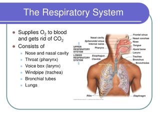
The Respiratory System. Supplies O 2 to blood and gets rid of CO 2 Consists of Nose and nasal cavity Throat (pharynx) Voice box (larynx) Windpipe (trachea) Bronchial tubes Lungs. Functions of Respiratory System. Cleaning and humidifying air Ventilation Gas exchange Gas transport

The Respiratory System
E N D
Presentation Transcript
The Respiratory System • Supplies O2 to blood and gets rid of CO2 • Consists of • Nose and nasal cavity • Throat (pharynx) • Voice box (larynx) • Windpipe (trachea) • Bronchial tubes • Lungs
Functions of Respiratory System • Cleaning and humidifying air • Ventilation • Gas exchange • Gas transport • Smell • Speech
Airways –respiratory tree Alveolar Sac
Pulmonary Lobule with Alveolar Sac Alveolar Sac in Pulmonary Lobules
Pulmonary Lobule • Lung lobules are smaller compartments within bronchopulmonary segments of lobes • Consist of • Terminal bronchiole supplies air to lobule • Each terminal bronchiole divides into several respiratory bronchioles • Respiratory bronchioles divide into alveolar ducts • Alveolar ducts supply air to alveolar (air) sacs • Each sac composed of smaller alveoli(us) • Blood supply – lab model terminal bronchiole respiratory bronchiole alveolar duct alveolar sac alveolus
Details of Alveolar Structure Pneumocyte III (macrophage) Pneumocyte I (alveolar cell) Lung capillary Pneumocyte II (surfactant cell) Respiratory membrane
Respiratory Membrane (RM) CO2 O2
Respiratory Physiology • Ventilation • Control of Ventilation • Gas Exchange • Gas Transport
Ventilation • Basic concepts and definitions • Breathing in is called inhalation or inspiration • Breathing out is called exhalation or expiration
Ventilation • Inhalation occurs when pressure inside lungs (intrapulmonary) pressure) becomes less than (negative to) atmospheric pressure • Contraction of diaphragm and rib muscles (ext. intercostals) enlarges chest and reduces intrapulmonary pressure to below atmospheric pressure • Air drafts into lungs
Ventilation • Exhalation occurs when pressure inside lungs becomes greater than (positive to) atmospheric pressure • Diaphragm and rib muscles (ext. Intercostals) relax • Chest gets smaller • Elastic recoil of chest and lungs, and contraction of certain muscles create intrapulmonary (lung) pressure greater than (positive to) atmospheric pressure • Air is pushed (squeezed) out of lungs
Ventilation Explained by Boyle’s Law • Boyles’s Law • As size of container increases, pressure inside decreases • As the size of container decreases, pressure inside is increases
Resting Ventilation-Eupnea • Inhalation is active • Contraction of diaphragm and external intercostal muscles expands chest and lungs, and decreases intrapulmonary pressure • Exhalation is passive • Relaxation of diaphragm and external intercostal muscles decrease chest volume, followed by elastic recoil of lungs increase intrapulmonary pressure to above atmospheric pressure
Forced Ventilation • Forced inhalation and exhalation • Must be larger change in chest and lung volumes resulting in larger changes in intrapulmonary pressure • Other muscles become involved resulting in deeper inhalation followed by more forceful exhalation • Muscles not only expand chest on inhalation, they compress chest on exhalation
Respiratory Volumes and Capacities • Volume is one measure of quantity of air • Capacity is sum of two or more volumes • Spirometer or respirometer device for measuring volumes and capacities • Record called spirogram
Ventilation Rates and Volumes • Ventilation rate is number of breaths per minute • Resting ventilation rate is12-18 breaths per minute • Tidal volume (TV) or (VT) is amount of air in one breath • Resting tidal volume is around 500 mL
Ventilation Rates and Volumes • Respiratory Minute Volume • If rate = 12 breaths per minute • And Tidal volume (VT) = 500 mL per minute • Then respiratory minute volume =
Ventilation Rates and Volumes • If resting tidal volume (VT) = 500 mL, then • Alveolar Ventilation (VA) • 350 mL reaches alveoli • Participates in gas exchange • Anatomic Dead Space or Air (VD) • 150 mL remains in conducting airways above alveoli • This air does not participate in gas exchange • Is mixture of fresh inhaled air and air to be exhaled
More Volumes and Capacities • Inspiratory Reserve Volume (IRV) • Air inhaled in addition to tidal volume • Might be around 3300 mL • Provides more inhaled air when active • Expiratory Reserve Volume (ERV) • Air exhaled in addition to tidal volume • Might be around 1000 mL
More Volumes and Capacities • Residual Volume (RV) • Amount of air that cannot be exhaled • Lungs must remain inflated • Might be around 1200 mL • Vital Capacity (VC) • Maximum amount of air inhaled and exhaled in one respiratory cycle • VC = VT + IRV + ERV • What is VC for the volumes we just used?
Control of Ventilation • Respiratory Centers of Medulla Oblongata • Dorsal Respiratory Group (DRG) • Innervates diaphragm and external intercostal (rib) muscles • Neurons control basic rhythm of resting ventilation (eupnea) • Inspiration for 2 seconds, expiration for 3 • Ventral Respiratory Group (VRG) • Inactive during most quiet breathing most active during forced ventilation • Innervates more muscles used in forced ventilation
Control of Ventilation • Neurons in the Pons coordinate transition between inhalation and exhalation • Apneustic area stimulates DRG • results in prolonged inhalation • Pneumotaxic area • inhibits apneustic center • causes passive and active exhalation • Helps to prevent over-inflation of lungs
Respiratory Reflexes • Chemoreceptor Reflexes • Sensitive to CO2, pH, and O2 in blood and CSF • Increase in CO2 excites chemoreceptors in carotid arteries, aorta and medulla oblongata and breathing rate increases – has greatest chemical effect on breathing rate • pH stimulates chemoreceptors causing increase in breathing rate • Dramatic decrease in O2 such as at high altitude stimulate chemoreceptors causing increased breathing rate
Respiratory Reflexes • Proprioceptors in muscles and Joints • Movement stimulates these receptors • Cause rapid increase in breathing rate with increased activity
Respiratory Reflexes • Stretch receptors in lungs • Inflation Reflex • Inhibits inhalation • Prevents over-inflation (Hering-Breuer reflex) • Deflation Reflex • Inhibits exhalation and starts inhalation • Only active during forced (active) ventilation
Respiratory Reflexes • Protective Reflexes • Coughing and Sneezing • Sneezing caused by irritants in nasal cavity • Coughing caused by irritants in larynx or bronchi • Both involve: • Apnea followed by forceful expulsion of irritant • Air speed may approach 100 mph in trachea • Laryngeal spasms • Caused by irritant or foreign object • Closes airway temporarily to protect airways
Gas Exchange in Lungs – External Respiration • Diffusion of O2 from alveoli into blood • Diffusion of CO2 from the blood into alveoli • Diffusion across the extremely thin (0.5m) respiratory membrane from higher to lower concentrations of gases CO2 O2 O2 CO2
Gas Exchange in Tissues – Internal Respiration • Diffusion of O2 from blood into tissues • Diffusion of CO2 from tissues into blood Cell CO2 O2 IF O2 CO2 Tissue Capillary
Gas Exchange Diagram External Respiration in Lungs Internal Respiration in Tissues Left side of Heart O2 O2 CO2 CO2 O2 Oxygenated Blood Alveolus CO2 Lung Capillaries O2 Tissue Capillaries CO2 O2 O2 CO2 Right side of Heart CO2 Deoxygenated Blood Tissue Cell
Measurements of Oxygen and Carbon Dioxide Concentrations • Measurements in Percentage • Atmospheric Air • Oxygen = 20.9% • Carbon dioxide = 0.04% • Alveolar Air • Oxygen = 13.6% • Carbon dioxide = 5.2% • Why the differences? Look for explanation on page 852 & 853 in textbook.
Measurements of Oxygen and Carbon Dioxide Concentrations • Measurements in Partial Pressures • Based on Dalton’s Law of Partial Pressure which states that in a mixture of gases, the total pressure is equal to the addition (sum) of the individual gas pressures • These individual pressures are partial pressures • Symbol for the partial pressure of a gas is Pg where g stands for the specific gas, so • PO2 stands for partial pressure of oxygen • PCO2 stands for partial pressure of carbon dioxide
Calculations of Partial Pressures • Standard atmospheric pressure at sea level= 760 mmHg • Atmospheric PO2 = • Atmospheric PCO2 = • Alveolar PO2 = • Alveolar PCO2 = 159 mmHg 0.3 mmHg 100 mmHg 40 mmHg
Partial pressures in Lungs- External Respiration • Partial pressure (concentration) of oxygen in alveolar air is greater than in deoxygenated blood, so O2 diffuses from alveolar air into blood • Partial pressure (concentration) of carbon dioxidein deoxygenated blood is greater than in alveolar air, so CO2 diffuses from blood into alveoli
Partial pressures in Tissues- Internal Respiration • Partial pressure (concentration) of oxygen in oxygenated blood greater than in tissues, so O2 diffuses from blood into tissues • Partial pressure (concentration) of carbon dioxidein tissues greater than in oxygenated blood, so CO2 diffuses from tissues into blood
Question Which of the following has a higher PO2? (A) Oxygenated blood (B) Alveolar air (C) Deoxygenated blood (D) Tissues
Question • Why does the PO2 of lung deoxygenated blood rise from 40 mmHg to 95 mmHg?
Henry’s Law • Formally stated • The amount of gas that will dissolve in a liquid is proportional to the partial pressure of the gas, and the solubility of the gas in the liquid • What is the effect of temperature? • More oxygen in the air, results in more oxygen in the blood
Henry’s Law • What is an “everyday” example of Henry’s law? • How does Henry’s law account for the effects of high altitude? • What is a clinical applications of Henry’s law?
Question • Why is tissue (cell) PO2 less than that of oxygenated blood?
Question Why is alveolar PO2 at 10,000 ft only 59 mmHg?


