Download
1 / 13
150 likes | 387 Vues
Evaluation of the Mature Cataract. Baxter McLendon MD FACS Clinical Professor of Ophthalmology Medical University of South Carolina Charleston, South Carolina USA. Many ocular surveys have reported poor vision post cataract surgery as a leading cause of blindness. WHY IS THIS ?

E N D
Evaluation of theMature Cataract Baxter McLendon MD FACS Clinical Professor of Ophthalmology Medical University of South Carolina Charleston, South Carolina USA
Many ocular surveys have reported poor vision post cataract surgery as a leading cause of blindness. WHY IS THIS ? Poor pre-op evaluation/ selection probably accounts for some of this blindness.
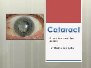
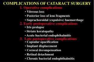
The Problem • No view of posterior pole with mature (white or brown) cataract • All mature cataracts should NOT automatically under go surgery
Pre-op Slit Lamp Evaluation • IOP 08 - 22 mm Hg • No bullous keratopathy • Red reflex present • No anterior uveitis / cells in A. C. • No vitreous in A.C. / no rubeosis • No phacodonesis / iridodonesis • No traumatic mydriasis • No iris retroillumination defect • No pupillary ruff abnormality/ defect
B Scan (Ultrasound) May Help Diagnose • Retinal Detachment • Vitreous Opacity [ blood, etc. ] • Posterior Pole Tumor • Choroidal Detachment • Posterior Scleral Thickening • Staphyloma
Three Step Clinical Test • Takes 90 seconds / easy • Can be done in or outside the clinic setting especially when B Scan is not available • No additional cost/ travel for patient • Helps determine post-op PROGNOSIS
#1 Check Pupils • Dim the lights • Have patient look (fixate) at end of room • Brisk or sluggish pupil compared to other eye • Afferent pupillary defect (Marcus Gunn)
#2 Color Perception • Only one chance to do test correctly. • Don’t let pt see test color pre - testing • Cover/occlude completely the other eye • Dim the lights (illumination) • “ What color is this light?”
#2 Color Perception • Check color red: red, pink, orange, or yellow are all acceptable answers • Check color blue: blue or green are acceptable [ correct ]
If the patient can NOT distinguish colors do NOT operate on mature cataract
#3 Light Projection • Dilate mature cataract pupil • Have patient look at the light. “ Where is the light“. “Follow the light “ • Have pt. point in the direction of the light [ up, down, right, left, etc. ]
Three Step Clinical Test • Checking pupils: optic nerve • Checking color perception: macula and optic nerve • Light projection: retinal detachment, diffuse C-R, and advanced glaucoma
In Conclusion If all three tests are normal, then the prognosis is usually quite good for improved vision after mature cataract surgery. This is quick, easy, and in 90 seconds you can determine if the patient is a good candidate or not for mature cataract extraction