CATARACT
CATARACT. Done by : Raya Marji. Objectives :. The Lens : anatomy and histology Defenition and epidemiology Symptoms and signs DDx of gradual painless loss of vision Classification Treatment Congenital cataract. The lens.

CATARACT
E N D
Presentation Transcript
CATARACT Done by : Raya Marji
Objectives : • The Lens : anatomy and histology • Defenition and epidemiology • Symptoms and signs • DDx of gradual painless loss of vision • Classification • Treatment • Congenital cataract
The lens • Biconvex, transparent ,purely epithelial structure without any nerves or blood vessels. • Lies in the posterior chamber of the eye between the posterior surface of the iris and the vitreous body. • Radially arranged zonule fibersconnect between ciliary body and lens capsule .These fibers hold the lens in position and transmit changes in the ciliary muscle allowing the lens to change its shape and refractive power . • Functions : one of the essential refractive media of the eye and focuses incident rays of light on the retina. • The lens is nourished by diffusion from the aqueous humor.
Histology: • Capsule : replicated basal lamina, formed from the basement membrane of the epithelium. • Subcapsular epithelium (simple cuboidal). • lens fibre production. • Synthesis of crystallins and membrane proteins • transpor ions and water 3. Lens fibers Lens fibres are nucleated in the cortex . As new lens fibres are added to the periphery of the cortex, lens fibres located deeper in the cortex loose their nuclei and become part of the harder nucleus of the lens.
Age-related changes • The water content of the lens is normally stable and in equilibrium with the surrounding aqueous humor. • The water content of the lens decreases with age, whereas the content of insoluble lens proteins (albuminoid) increases. The lens becomes harder, less elastic (Loss of accommodation), and less transparent. • A decrease in the transparency of the lens with age is as unavoidable. • The nucleus of the lens becomes sclerosed and slightly yellowish .
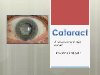
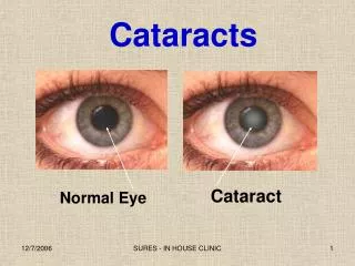
Cataract • Opacification ( clouding ) of the lens inside the eye which leads to a decrease in vision . • Is present when the transparency of the lens is reduced to the point that the patient’s vision is impaired. • The commonest cause of treatable reversible decrease in vision in the world !
When eyes work properly: Light passes through the cornea and the pupil to the lens. The lens focuses light & producing clear, sharp images on the retina.As a cataract develops, the lens becomes clouded, which scatters the light and prevents a sharply defined image from reaching retina. As a result, vision becomes blurred.
Epidemiology • 60% of those aged 65 to 74 years, and by 91% of those aged 75 to 85 years. • Frequency rates of cataracts are on the rise. • More in females than males
symptoms Painless , gradual loss of vision Glare Change in refractive error . Altered colours perception Haloes may be observed around lights In certain types of cataracts, double vision may be noted
Signs • Reduced acuity. • Appears dim against red reflex when the eye is examined with direct ophthalmoscope . • Reduced contrast . • Slit lamp examination allows the cataract to be examined in detail and the exact site of the opacity can be identified . • Severe dense cataract cause a white pupil.
-Normal red reflex is : Diffuse bright red -Causes of dim red reflex : *anything that interfere with the passage of light from cornea to retina 1-corneal ulcer , keratitis . 2-Hyphemia ( Anterior champers). 3-Cataract 4- vitreous hemorrhage . 5- NOT GLAUCOMA .
Red reflex SLIT LAMP EXAMINATION
Painless Gradual loss of vision DDX: • Refractive error • Cataract • Age related macular degeneration ( AMD ) • Glaucoma (open angle glaucoma) • Diabetic maculopathhy (not retinopathy) .
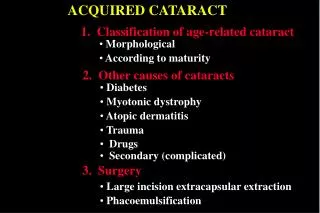
Classification of cataract • Cataracts can be classified by the cause of the cataract , anatomical location within the lens, degree of clouding of the lens . • Cause : acquired vs congenital. • Anatomical : nuclear , cortical , sub-capsular. • Degree of clouding (degree of loss of the normal transparency) : Immature , mature , hyper-mature .
According to the cause : • Acquired 1- age-related ( more than 90%) 2- ocular conditions : high myopia, trauma, uveitis , intraocular tumor , Previous ocular surgery * Topical medications : steroid eye drops . 3- systemic conditions : DM , atopic dermatitis , Myotonic dystrophy , radiation *systemic medications : steroid • Congenital • Non-systemic association : Sporadic , Autosomal dominant, Recessive,X-linked • Systemic Associations : metabolic associations : Galactoasemia intrauterine infections : Congenital rubella , Toxoplasmosis , Varicella chromosomal abnormalities : Down , Edwards syndrome .
Age –related(Senile) cataract Most common . Lens proteins denature and degrade over time , and due to cumulative exposure to environmental influences such as smoking , UV radiation . Classified according to: • Morphology • Nuclear • Cortical • Subcapsular • Maturity{degree of cloudiness } • Immature Cataract • Mature Cataract • Hypermature Cataract
Nuclear cataract • Most common type. • Nuclear cataract starts as an exaggeration of the normal ageing changes involving the lens nucleus (Age related lens hardening ) . • Nuclear Cataracts begin with gradual hardening and yellowing of the center of lens ,called the nucleus. This hardening gradually expands to the other layers of the lens & develop very slowly. • It is often associated with myopia (nearsighted) due to an increase in the refractive index of the nucleus, and increased spherical deviation . In early stages some elderly patients may consequently be able to read without glasses again ('second sight of the aged’). • Unfortunately, this so-called 2nd sight is temporary and disappears as the lens gradually turns more densely yellow & further clouds vision. • As the cataract progresses, the lens may even turn brown, black discoloration of the entire lens (black cataract)may occur too.Advanced discoloration can lead to difficulty distinguishing between shades of blue & purple (contrast is affected ). • This type of cataract is best assessed with oblique slit-lamp biomicroscopy and not by retroillumination .
Nuclear cataract: The nucleus of the lens has a yellowish brown color due to DEPOSITION OF UROCHROME PIGMENT..
Yellow Brown Black
Cortical cataract • Occur on the outer edge of the lens (cortex) , and may involve the anterior, posterior or equatorial part of cortex. • The opacities start as clefts and vacuoles between lens fibers due to hydration of the cortex. Subsequent opacification results in typical whitish wedge-shaped opacities or streaks , often initially in the inferonasalquadrant of the lens . • The streaks extend to the center and interfere with light passing through the center of the lens. • It’s slowly progresses, but more rapid than nuclear . • CC by increased water content (swollen lens ) unlike nuclear cataract which is cc by hardening. • Tend to have acquired hyperopia in contrast to patients with nuclear cataracts, who tend to be myopic • Patients with cortical opacities frequently complain of glare due to light scattering. . • Visual acuity may temporarily improve during the course of the disease. This is due to a stenopeic effect as light passes through a clear area between two radial opacities
Subcapsular cataract • Occur just under the capsule of the lens. • Starts as a small, opaque area. • It interferes with reading vision. • Reduces vision in bright light ( Glare) • Causes glare or halos around lights at night.
Anterior subcapsular cataract • Definition : lies directly under the anterior lens capsule and is associated with fibrous metaplasia of the lens epithelium. • Incidence/Prevalence: Anterior subcapsular cataract is less common than other types of cataract. The prevalence is about 1/5 that of posterior subcapsular opacities . • Associations : atopic dermatitis, steroid ,inflammation, trauma . • Clinical Findings: A focal star-shaped or irregular opacity beneath the anterior capsule as seen with a slit beam.
Posterior Subcapsular Cataracts • Posterior subcapsular opacity lies just in front of the posterior capsule and has a granular clusters or plaque-like appearance on oblique slit-lamp biomicroscopy and spreads (expands )peripherally in a disk-like pattern. • Due to its location at the nodal point (point where the light converges, 2nd refraction point) of the eye, a posterior subcapsular opacity has a more profound effect on vision than a comparable nuclear or cortical cataract as it leads to early, rapid, and severe loss of visual acuity . Near vision is frequently impaired more than distance vision. • Patients are particularly troubled under conditions of miosis, such as produced by headlights of oncoming cars and bright sunlight. • Dilating drops useful in this type by keeping the pupils large and thus allow more light into the eye.
Done with Morphological Classification Lets go to Maturity classification
An immature cataract has some transparent protein, but with a mature cataract, all the lens protein is opaque. In a hypermature or Morgagnian cataract, the lens proteins have become liquid.
Immature cuneiform senile cortical cataractLens is partially opaque
Mature Cataract • The lens is diffusely white due to complete opacificationof the cortex. • Lens appears pearly white. • Vision reduced to just perception of light • Iris shadow is not seen • lens with a mature cataract can swelland acquire a silky glitter . The increasing thickness of the lens increases the resistance of aqueous humor flow from post to ant. champer at mid dilated stage of the pupil and with it the risk of angle closure glaucoma.
Mature cataract: • There is diffuse, complete opacification of the lens. A brownish nucleus is faintly visible posterior to the cortical layer. • Interior of the eye is no longer visible. • Visual acuity is reduced to perception of light and dark.
Hypermature Cataract • Occurs If a mature cataract progresses to the point of complete liquification of the cortex, the dense brown nucleus will subside within the capsule. • This may take any of two forms: • Liquefactive/Morgagnian Type • Sclerotic Cataract
Liquefactive/Morgagnian Type • When the lens capsule becomes permeable for liquified lens substances, it will lose volume due to leakage. The capsule will become wrinkled and shrunken . The escaping lens proteins will cause intraocular irritation and attract macrophages that then cause congestion of the trabecular network (Secondary open angle glaucoma). • Cortex undergoes auto-lytic liquefaction and turns uniformly milky white. • The nucleus loses support and sinks inferiorly .
Cataract maturity. (A) Mature cataract; (B) hypermature cataract with wrinkling of the anterior capsule; (C) Morgagnian cataract with liquefaction of the cortex and inferior sinking of the nucleus; (D) total liquefaction and absorption of the cortex with inferior sinking of the lens
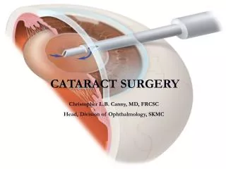
Treatment • Glasses: are not used as line of tt ,however; after cataract surgery the capsule will be defected and the patient will lose the accommodation ability so he will need glasses for near vision • Surgical removal : is the gold standerad . • Surgical techniques • Phacoemulsification method. • Extracapsular method. • Intracapsular method not used anymore , not required.
Pre-op assesments • General health evaluation including blood pressure check • Assessment of patients’ ability to co-operate with the procedure and lie reasonably flat during surgery • Instruction on eye drop instillation • The eyes should have a normal pressure, or any pre-existing glaucoma should be adequately controlled on medications. • An operating microscope is needed, in order to reach the lens, a small corneal incision is made close to the limbusfor the phaco-probe. • It is important to appreciate anterior chamber depth and to keep all instruments away from the corneal endothelium in the plane of the iris.
Phacoemulsification: Phacoemulsification is the most widely used cataract surgery. This procedure uses ultrasonic energy to emulsify the cataract lens. Anaesthetic - The eye is numbed with either a subtenon injection around the eye or using simple eye drops. Cornealincision - Two cuts are made through the clear cornea to allow insertion of instruments into the eye. Capsulorhexis - A needle or small pair of forceps is used to create a circular hole in the ant. capsule in which the lens sits. Phacoemulsification - A handheld probe is used to break up and emulsify the lens into liquid using the energy of ultrasound waves. The resulting 'emulsion' is sucked away.
Irrigation and aspiration - The cortex, which is the soft outer layer of the cataract, is aspirated or sucked away. Fluid removed is continually replaced with a saline solution to prevent collapse of the structure of the anterior chamber (the front part of the eye). • Lens insertion - A plastic, foldable lens is inserted into the capsular bag that formerly contained the natural lens.
. Phacoemulsification in cataract surgery involves insertion of a tiny, hollowed tip that uses high frequency (ultrasonic) vibrations to "break up" the eye's cloudy lens (cataract). The same tip is used to suction out the lens
Extra-capsular Cataract Extraction (ECCE) Extracapsular cataract extraction (ECCE) consists of removing the lens manually, but leaving the majority of the capsule intact. The lens is expressed through a 10– to 12-mm incision which is closed with sutures at the end of surgery. ECCE is less frequently performed than phacoemulsification, but can be useful when dealing with very hard cataracts or other situations where emulsification is problematic
Extra-capsular Cataract Extraction (ECCE) • The nucleus and the cortex is removed out of the capsule leaving behind: • Intact posterior capsule • Peripheral part of the anterior capsule • Zonules. • This method: • Provides support of placement of IOL • Prevents vitreous from bulging forwards • Acts as a barrier between anterior and posterior segment. • All this results in decreasing the incidence of complications.
Postoperative care after cataract surgery • Steroid drops (inflammation) • Antibiotic drops (infection) • Avoid • Very strenuous exertion (rise the pressure in the eyeball) • Ocular trauma.
Complications of cataract surgery • Infective endophthalmitis • Rare1-2/1000 but can cause permanent severe reduction of vision. • Most cases within two weeks of surgery. • Typically patients present with a short history of a reduction in their vision and a red painful eye. • This is an ophthalmic emergency. • Low grade infection with pathogen such as Propionibacterium species can lead patients to present several weeks after initial surgery with a refractory uveitis (staph epidermidis ) 3-prohylaxis: inter-vitreal steroids, IV antibiotics 4-management: vitrectomy • Suprachoroidalhaemorrhage. • Severe intraoperative bleeding can lead to serious and permanentreduction in vision.
Uveitis • Postoperative inflammation is more common in certain types of eyes for example in patients with diabetes or previous ocular inflammatory disease. • Ocular perforation. • Postoperative refractive error • Most operations aim to leave the patient emmetropic or slightly myopic, but in rare cases biometric errors can occur or an intraocular lens of incorrect power is used. • Posterior capsular rupture and vitreous loss • If the very delicate capsular bag is damaged during surgery or the fine ligaments (zonule) suspending the lens are weak (for example, in pseudoexfoliation syndrome), then the vitreous gel may prolapse into the anterior chamber. This complication may mean that an intraocular lens cannot be inserted at the time of surgery. Patients are also at increased risk of postoperative retinal detachment.