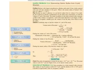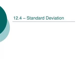
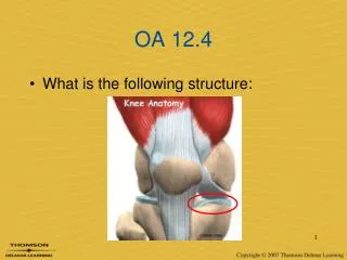
OA 12.4
OA 12.4. What is the following structure:. Chapter 18 (pp 471-486). T he Knee. Anatomy. Objectives. Identify… The bones of the knee The ligaments of the knee The muscles of the knee The tendons of the knee The blood vessels & nerves of the knee Other structures. Bones. Femur (1)

OA 12.4
E N D
Presentation Transcript
OA 12.4 • What is the following structure:
Chapter 18 (pp 471-486) The Knee
Objectives Identify… • The bones of the knee • The ligaments of the knee • The muscles of the knee • The tendons of the knee • The blood vessels & nerves of the knee • Other structures
Bones • Femur (1) • Tibia (3) • Fibula (4) • Patella (2)
Bones: landmarks • The proximal end of the tibia is called the plateau • Attachment site for ACL, PCL, Meniscus • The proximal end of the fibula is called the head (apex) • Attachment site for LCL
Bones: landmarks • The tibial tuberosity is the bony outgrowth on the anterior aspect of the tibia • Attachment site for patellar tendon
Bones: landmarks • The distal end of the femur is has two condyles, and just superior to those are epicondyles • Medial = MCL & lateral = LCL
Bones • The patella is the largest sesamoidbone in the body • Housed within quadriceps/ patellar tendon
Menisci • The knee contains two meniscus– medial & lateral • Medial = C shaped • Lateral = O shaped
Menisci • The meniscus is only partly vascularized (only part receives blood flow) • Known as the vascularor avascular zone
Menisci • Fibrocartilage • Deepens the joint • Increases stability • Absorbs shock • Lubricates the joint
Ligaments • There are four main stabilizing ligaments in the knee • Two cruciates (crossing) • Two collaterals(on the sides) • Anterior cruciate ligament • Posterior cruciate ligament • Medial collateral ligament • Lateral collateral ligament
Ligaments ACL PCL Posterior-lateral tibia to medial condyle of femur Prevents internal rotation of the tibia & guides the knee during flexion • Anterior-medial tibia to lateral condyle of femur • Three bands • Prevents internal rotation of the tibia & anterior translation
Ligaments MCL LCL Lateral epicondyle of the femur to head of the fibula Protects against varusforces • Medial epicondyle of femur to tibia • Protects the knee from valgusforces & external rotation of the tibia
Articulations • The knee is comprised of three articulations • Tibiofemoral= tibia & femur • Tibiofibular= proximal tibia & fibula • Patellofemoral= patella & femur
Muscles & tendons • Anterior aspect – extend the knee (and flex the hip) • Quadriceps femoris group • Vastusmedialis • Vastusintermedius • Vastuslateralis • Rectus femoris • Sartorius*
Muscles & tendons • The quadriceps group forms the quadriceps tendon • Pulls on the patella to extend the knee
Muscles & tendons • Posterior aspect – flex the knee* (and extend the hip) • Hamstrings group • Semitendinosis • Senimembranosis • Biceps femoris • Gastrocnemius • Plantaris • Popliteus
Muscles & tendons • Three muscles join to form the pes anserine group, which insert on the anterior tibia • Sartorius, Gracilis, SemiTendinosus • Acronym: SGT. (sargeant) • Knee flexors
Muscles & tendons • The lateral aspect of the knee is controlled/stabilized by the iliotibial band (IT Band (8)) • Connective tissue that attaches the TFL muscle to the knee at Gerdy’s Tubercle (7)
Other structures • The posterior aspect of the knee is known as the popliteal fossa • Popliteus muscle • Popliteal tendon • Popliteal artery • Popliteal nerve
Other structures • Bursae – as many as two dozen around the knee • Suprapatellar (A&B) • Prepatellar (C&D) • Infrapatellar(F, G, H) • Pes anserine (I)
Other structures • Fat pads – exist to cushion the knee
Other structures • Nerve supply • Tibial nerve • Common peroneal nerve • Femoral nerve • Located on… • Posterior aspect • Lateral aspect • Anterior-Medial aspect
Other structures • Blood supply • Femoral artery (2) popliteal artery (3) • Pulse is felt at the popliteal artery, in the popliteal fossa
OA 12.9 If an athlete came to you complaining of knee pain, how would you address them? • What questions would you ask to gather clues about what is going on? • What are some relevant observations to make regarding their body?
Objectives Identify… • Pertinent information to gather during a knee evaluation • Important observations to make during a knee evaluation ???
Introduction • Must understand anatomy& biomechanics • Examination process is on-going • Initial rehab RTP • Must be systematic and methodical • Must understand differential diagnosis (DDx) • Options that a specific injury could be • Pathologies often have similar S&S • Rule out emergency situations quickly • If unsure, err on side of caution
History • Start with generic history questions • Chief complaint • Age • Occupation / sport / position etc. • General healthcondition • Activity level • Medications
History • History of previous injuries • What happened? • Who did you see? • What did they tell you? • How long were you out? • Has it fully resolved?
History • Mechanism of injury • How did it happen? • Tension = sprain; fracture; strain • Torsion = sprain; fracture • Compression = contusion; fracture • Shear = fracture; sprain • Bending = fracture
History • Mechanism of Injury – Knee specific • Hyperextension • Valgus force – lateral medial • Varus force – medial lateral • Compression • Internal/External rotation of the tibia • Overuse
Hyperextension Internal rotation of the tibia
Valgus Force Normal landing
History • Ask these questions regarding PAIN • P-rovocation – what causes it? what makes it better? • Q-uality – what does it feel like? neurological symptoms? • R-egion – where does it hurt? can you point w/one finger? • S-everity – how bad does it hurt? (1-10) • T-iming – when does it hurt? how long?
History • Sounds & sensations • Did you hear any sounds? Did you hear any pops, crackles, snaps, clicking? • What could this indicate??? • Did you feelanything unusual?
History • Specific to the KNEE • Previous history = instability, LAXITY of the ligaments • Mechanism of injury = Hyperextension, Valgus, Varus, Chronic overuse, etc. • Location of pain – Determineswhat is injured • Any changes in activity,footwear, or training surfaces
History • Specific to the KNEE • Did the knee collapseor give out? • Could you bend/straighten your knee afterwards? • Did it begin to swell? • Is there any clicking or grinding sounds? • Is there pain with walking? • Pain with sitting for long periods of time? • Pain going up/down stairs?
Observation • When does this begin? • Compare each side bilaterally to identify what is normal for that person We look for: • Deformity, asymmetry, edema, ecchymosis
Observation For the knee: • Gait • Half squatting • Going up/down stairs • Leg alignment • Genu valgum–knocked knee’d • Genu varum– bow-legged • Genu recurvatum– Hyperextension • Patella alta– high patella • Patella baja– low patella • Squint eye patella – points medially • Frog eye patella –points laterally