Download
1 / 45
450 likes | 779 Vues
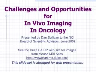
Challenges and Opportunities for In Vivo Imaging In Oncology. Presented by Dan Sullivan to the NCI Board of Scientific Advisors, June 2002. See the Duke SAIRP web site for images from Mouse MRI Atlas http://wwwcivm.mc.duke.edu/. This slide set is abridged for web presentation.

E N D
Challenges and Opportunitiesfor In Vivo ImagingIn Oncology Presented by Dan Sullivan to the NCI Board of Scientific Advisors, June 2002 See the Duke SAIRP web site for images from Mouse MRI Atlas http://wwwcivm.mc.duke.edu/ This slide set is abridged for web presentation.
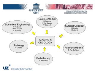
In Vivo Imaging For Oncology Devices (Hardware &Software) Laboratory Methodologies Study biology or interventions in animal models or humans (e.g., functional, in vivo genomics/proteomics; drug development) • Clinical Methodologies • Screening • Diagnosis • Staging • Prognosis • Therapy Monitoring • Image-guided Rx Molecular Probes; Contrast Agents Image Exploitation; Informatics
Outline • Overview • Look back (portfolio review) • Clinical and Laboratory imaging issues • Molecular Imaging • Image-guided interventions • Conclusion
Imaging in Clinical Trials • ACRIN – more later • Rigorous methodology • Two U01’s will not be renewed • Additional venues for diagnostic trials: e.g., ACOSOG; R01’s; R33’s
Imaging in Clinical Trials • More integration of imaging studies in CTEP trials – especially in the neoadjuvant setting • Inter-group Imaging Council • Imaging Cores in Cancer Centers • Access to imaging resources is inadequate
Monitoring Response to Therapy • RECIST Criteria -- good start; volumetric would be better. • Need Functional Response Indicators – “molecular imaging”. • Generic, “downstream” indicator vs. specific, targeted probe?
Chemotherapy Response by MRI & MRS 1 wk pre-Tx 76 cc Day 1 AC x1 79 cc Day 42 AC x3 26 cc Day 70 AC x4 25 cc Day 112 taxol x2 11 cc Day 178 taxol x4 6 cc 486 593 267 79 481 595 partial response to AC, regrowth on taxol final pathology - viable IDC and extensive DCIS Univ. of Minnesota
Molecular Imaging Targets/Probes MAb,Fragments Receptor Mapping Hormones Drugs and Ligands Peptides Accumulation via Phosphorylation [18F]FDG Enzyme Activity: Inhibition, Conc., Synthesis Internalization glut 4 Hexokinase Accumulation via DNA-Synthesis DNA Accumulation via AA Transport or Protein Synthesis AAT mRNA Oligonucleotides mRNA Binding mRNA DNA Reporter Gene Reporter Probe
FDG-PET Monitoring Response to STI571 in GIST Baseline 24 hrs 7 days 2 mos 5.5 mos Dana-Farber Cancer Institute
2 mm In vivo imaging of protease (Cathepsin B) activity Light image NIRF image Human LX1 small cell lung tumor 10 nmole C-PGC, 24 hr Nature Biotech; 1999;17:375-378 Weissleder, Nature Biotech, 1999
A Nanoscale, Targeted Liposome Targeting MAb or ligand High affinity metal binding Gd encapsulation Stanford Univ.
Phase 1, 2 Clinical Trials MMPI and Cancer: Trials and Tribulations, Coussens, Science 2002
Targeted Agent Trials: • Problems: • Selecting patients • Biologic endpoint assessment • Examples: • Matrix metaloproteinase inhibitors • Anti-angiogenesis therapies • 17-AAG (HSP90 inhibitor)
PEG Image Therapy MSH CCK Enk. Construction ofMultimeric Ligands Gillies; U. AZ
Nano-engineered Optical Agents • Emission spectra can be “tuned” • [Nanotechnology: the creation of functional materials, devices and systems through control of matter at the scale of 1 to 100 nanometers, and the exploitation of novel properties and phenomena at the same scale.]
DCIDE Development of Clinical Imaging Drugs and Enhancers Purpose: Facilitate pre-clinical development of promising imaging agents by providing the resources needed for successful IND application.
Small Animal Imaging for Drug Development (Demonstration Project: BIP, DTP, CTEP) • anti-angiogenesis first model system; • NCI will supply the animals, drugs, and imaging protocol, to contractors; • tissues will be returned to NCI for standardized processing.
PET/CT • David Townsend, U Pitt. • NCI R01, 1993 • Now commercially available • ~ 150 expected to be sold by 2003 • Townsend has received R33 for second-generation • Higher-resolution, faster acquisition PET • Sub 4-minute scans
Quantitative whole body imaging in 5 min …………or less
PET/CT-Based IMRT: Cervical Cancer14 mm increase in axial extent of para-aortic lymph nodes
Imaging Modalities Physiology Molecular Anatomy CT US MRI PET Optical
FMT imaging of CAB in 9L glioma White light a) 1cm g) Excitation light 1cm b) (nM) (cm) 2 1 h) Emission light 1cm i) 1cm 0 Nature Med, 2002
108 106 104 30dB 20dB 102 Fluorescence strength (counts • sec-1 • mm-2) 10dB 100 Breast (40-70yrs) Breast (20-40yrs) Lung adult Muscle adult Brain adult 10-2 10-4 4 6 8 10 12 14 16 18 20 Depth (cm) Depth penetration in human tissues Ntziachristos, Ripoll, Weissleder. Optics Letters, 2002; 27:333-335
Whole-body Screening Now: • US – CT • Germany – MRI • Japan – FDG PET Future (US): • PET/CT? • MRI?
Skin Cancer Screening “Optical screening”; < 2% chance of AK4 Ca; Marked “Overdiagnosis” Rx is: “benign”, inexpensive, “Image-guided”.
MRI-guided, focused ultrasound therapy Ductal invasive breast cancer Treatment plan 80 ultrasound foci
0 40°C T MRI-guided, focused ultrasound therapy Temperature monitoring during therapy (SRTF, T1-w)
MRI-guided, focused ultrasound therapy Summary, properties • Noninvasive through intact skin • No scar • No anesthesia • No hospitalisation • Immediate Effect • Repeatable
Integrated Cancer Care Detection Diagnosis Treatment Detection Diagnosis Treatment data analysis sensing effector action judgment communication Unconventional Innovations Web Site:
UIP Awardees FY 99 U of Michigan, James Baker, M.D. Nano-scale based dendrimer devices U of Penn, Britton Chance, Ph.D. Near-infrared detector and contrast agents for molecular targets U of Alabama at Birmingham, David Curiel, M.D. Genetic approaches to tumor detection and intervention U of Cal at Davis, N.C. Luhmann, Jr.Ph.D. Compton light source for high-contrast X-rays; NASA Ames Research Center, Meyya Meyyapan, Ph.D. Carbon nanotube-based biosensor and prototype biosensor catheter
UIP Awardees FY 2000 U of Washington, Kirk W. Beach, M.D., Ph.D. Ultrasound detection of tissue pulsatility U of Pittsburgh, Daniel Farkas, Ph.D. Optical imaging platform for mesoscopic imaging U of Michigan, Raoul Kopelman, Ph.D. Dynamic nano-particles for detection and treatment Barnes-Jewish Hospital, Gregory M. Lanza, M.D., .Ph.D. Targeted Nanoparticle Emulsion for Molecular Imaging and Local Drug Delivery
In Vivo Imaging For Oncology Go back and review slide 2
Acknowledgements Ellen Feigal Laurence Clarke John Hoffman Gary Kelloff Edward Staab Houston Baker Barbara Croft Keyvan Farahani Barbara Galen Guoying Liu Anne Menkens Richard Reba Johnnie Smith James Tatum Manuel Torres Michael Vannier And Many Investigators.