IMAGING in ONCOLOGY
370 likes | 907 Vues
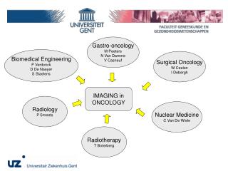
Gastro-oncology M Peeters N Van Damme V Casneuf. Biomedical Engineering P Verdonck D De Naeyer S Staelens. Surgical Oncology W Ceelen I Debergh. IMAGING in ONCOLOGY. Radiology P Smeets. Nuclear Medicine C Van De Wiele. Radiotherapy T Boterberg. Imaging of Angiogenesis.

IMAGING in ONCOLOGY
E N D
Presentation Transcript
Gastro-oncology M Peeters N Van Damme V Casneuf Biomedical Engineering P Verdonck D De Naeyer S Staelens Surgical Oncology W Ceelen I Debergh IMAGING in ONCOLOGY Radiology P Smeets Nuclear Medicine C Van De Wiele Radiotherapy T Boterberg
Imaging of Angiogenesis • Direct: In Vivo Microscopy; molecular probes • Indirect • Blood volume • Vessel surface area • Vessel permeability • Vessel ‘normalization’
Neoplastic vessels are morphologically and functionally deficient • Highly irregular and tortuous • Dependent on cell survival factors (VEGF) • Hyperpermeable • deficient pericyte coverage • absence of a basement membrane • deficient intercellular junctions • presence of cellular lacunae • vascular mimicry • Resulting in abnormal microenvironment: hypoxia, low pH, interstitial hypertension
Hyperpermeability of tumor vessels is important • Results in increased interstitial fluid pressure • Enhances macromolecular leakage • Permeability change is a surrogate marker of angiogenesis
The concept of vessel normalisation by anti-angiogenic therapy • Return to ‘normal’ phenotype by vascular pruning • Results in more efficient drug delivery by lowering IFP and restoring microvessel function paradoxical synergism of anti-angiogenesis agents and cytostatic drugs • Results in more efficient RT by enhanced oxygenation
Bevacizumab normalises rectal cancer microvessels Willett et al. NATURE MEDICINE VOLUME 10 | NUMBER 2 | FEBRUARY 2004
Known Effects of Radiotherapy on tumor vessels • Induces endothelial cell apoptosis • Induces EC ‘senescent’ phenotype • Stimulates production of endothelin • Effects on vascular permeability dependent on dose, fractionation, tissue type • Direct or bystander effect?
Apoptosis resistant TUNEL Garcia-Barros et al. Tumor Response to Radiotherapy (15 Gy) Regulated by Endothelial Cell Apoptosis. Science 2003
How does anti-angiogenic therapy increase radiotherapy efficacy? • Inhibits protective VEGF signaling from tumor EC • Enhances oxygenation • Reduces number of circulating EC and progenitor cells
Phase I trials combining neoadjuvant CRT with anti-angiogenic therapy in rectal cancer • Czito IJROBP 2007 • 11 patients, 50.4 Gy, capecitabine,oxaliplatin, and bevacizumab • Acceptable toxicity • 18% pCR; 27% microscopic disease • Willett JCO 2005 • 5 patients, 50.4 Gy, 5-FU, bevacizumab 10 mg/kg • 2 complete responses seen in patients with DLT
Functional imaging using Dynamic Contrast Enhanced MRI • Inject contrast agent bolus • Acquire a series of MR images over time with sufficient temporal resolution • Measure signal intensity changes as a consequence of leakage of CA from the vascular compartment into the interstitial space
DCE-MRI Data Analysis • Description of enhancement curve • Semi-quantitative approach: AUC, TTP, maximal enhancement • Pharmacokinetic compartmental models • Statistical models
Farmacokinetic models in DCE-MRI Tofts St Lawrence - Lee
Physiological significance of calculated endothelial transfer constant • Small MW contrast agent: flow >> permeability • Large MW contrast agent (preclinical): permeability >> flow
Oncology Imaging Research @ UZ Gent • Preclinical • DCE-MRI • Rat colorectal cancer model • Mouse pancreas cancer model • Molecular MRI • Sequence optimisation; simulation; modelling • In vivo flurescence microscopy • Hamster melanoma and pancreas cancer model • Human CRC model in athymic mouse • Clinical • DCE-MRI in metastatic CRC treated with folfox-bevacizumab • USPIO in advanced solid tumors In collaboration with research dept. of Guerbet SA
Effects of RT on neovascular permeability measured with DCE-MRI using P792 Ktrans (1/min) After RT Before RT Ceelen et al. Int J Radiat Oncol Biol Phys 2006
rhEPO prevents radiotherapy-induced reduction in neovascular permeability Ceelen et al. Br J Cancer 2007
Molecular imaging by targeting αvβ3 integrin Arg-Gly-Asp (RGD) peptide Beta 3 integrin
Pre 20 min Post Molecular imaging in HT29 tumors using a αvβ3 integrin targeted MR probe Debergh I, Van Damme N, Smeets P, Peeters M, Pattyn P, Ceelen W. Molecular imaging of tumor associated angiogenesis using P1227, a novel MRI contrast agent targeting avb3 integrin. Br J Surg 2008;95 Suppl 6: 13.
Collaboration with the biomedical engineering department • Modelling of the AIF in DCE-MRI (PhD student) • D De Naeyer et al. Modelling the arterial input function for P792 in dynamic contrast enhanced magnetic resonance imaging: concepts and validation. Submitted, MRM • Modelling interstitial fluid pressure and drug transport with computational fluid dynamics software (Fluent) based on DCE-MRI data (thesis student) • Hyperpolarized gas MRI; 19F MRI (hypoxia imaging) • Small animal nuclear imaging
CFD model of a solid tumor. Zhao et al. Microvascular Research 73 (2007) 224–236
Small animal nuclear imaging: installed hardware • Micro SPECT (spatial resolution 0.35 mm) • Micro CT (spatial resolution <15 µm), GE • Small animal PET • Dedicated radiopharmacy unit (Prof Devos)
DCE-MRI in patients with metastatic CRC using MS-325 Large liver metastasis
Aims of the project • Study interaction of RT with anti-angiogenic therapy • Verify phenomenon of ‘vascular normalization’ and its importance for therapy planning • Validate functional and molecular imaging as a tool to monitor therapy response • Validate CFD modelling of vascular normalization and drug transport
Methods • Athymic mouse model; human CRC cell line • AZD2171 (Cediranib, 6 mg/kg), RT, or both • Measurements and imaging • Non invasive: DCE-MRI; P1227 • Semi-invasive: IVM • Invasive: pO2, IFP • Modelling of IFP and drug transport using Fluent • Possibility to study nuclear molecular probes in parallel
AZD 2171 Wedge et al. Cancer Res 2005
Contact WP Ceelen, MD, PhD Ghent University Hospital Wim.ceelen@ugent.be M Peeters, MD, PhD Marc.peeters@ugent.be