Download
1 / 16
160 likes | 372 Vues
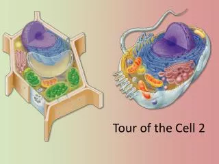
A Tour of the Cell, Part 2 Lecture 2, Part 2. Cytoskeleton. Network of fibers extending throughout the cytoplasm Three types: Microtubules Microfilaments Intermediate filaments. 1. Fig. 6.20. 2. Fig. 6.21. Cytoskeleton. Microtubules Hollow rods Protein: tubulin

E N D
Cytoskeleton Network of fibers extending throughout the cytoplasm Three types: Microtubules Microfilaments Intermediate filaments 1 Fig. 6.20
2 Fig. 6.21 Cytoskeleton Microtubules • Hollow rods • Protein: tubulin • Largest diameter (25 nm) • Actively assembled & disassembled • Function • Maintain cell shape • Compression-resisting • Cell division (centrioles) • Tracks for organelle movement • Cell motility (cilia & flagella) Table 6.3
Cilia & Flagella Microtubular containing extensions from a cell Flagella Typically one or two, long Undulating motion Cilia Typically many, short Back & forth motion Functions: Mobility of cell Movement of fluid past a cell Attachment (some cilia) Signal-receiving (specialized cilia) 3 Fig. 6.23
Microfilaments Solid rods Protein: actin Smallest diameter (7 nm) Actively assembled & disassembled Function Maintain cell shape Tension bearing Change cell shape Cell motility (pseudopodia) Cell division (cleavage furrow) Muscle contraction Cytoplasmic streaming 4 Cytoskeleton Table 6.3
Intermediate filaments Supercoiled cables Proteins: keratin family Intermediate & variable diameter (8-12 nm) Permanent structures Function: Maintain cell shape (tension bearing) Anchor nucleus, some organelles Forms nuclear lamina 5 Cytoskeleton Table 6.3
Extracellular Matrix (ECM): Animal Cells Collagen Glycoproteins (protein + carbohydrate) Proteoglycan complex Core protein with many carbohydrate chains 6 Fig. 6.30
7 Extracellular Matrix (ECM): Animal Cells • Fibronectin • Attaches ECM to integrins • Integrins • Cell surface receptor proteins Fig. 6.30
Tight junctions Continuous seal around cell Prevent fluid leakage Desmosomes Attach cells together Gap Junctions Cytoplasmic channels Proteins 8 Intercellular Junctions: Animal Cells Fig. 6.32
9 Cell Walls in Plants Cell walls • A protective layer external to the plasma membrane • Plants (also bacteria, archaea, fungi, some protists) Functions: • Protection • Maintain shape • Prevent excessive water uptake Fig. 6.28
10 Cell Walls in Plants • Matrix of cellulose, other polysaccharides & proteins • What is cellulose?
Carbohydrates Function Energy source used in cellular respiration to create ATP What are some sources of carbohydrates? Carbon skeleton “building block” for other organic molecules Cell identity Fibrous structural material Cellulose, chitin, peptidoglycan 11
Monomer small molecule Polymer large molecule made up of many smaller monomers Macromolecules “Big molecule” Synthesis & Breakdown of Polymers Dehydration reaction Hydrolysis 12 Fig. 5.2 Carbohydrates
Carbohydrates Monosaccharides Simple sugar (CH2O) E.g., glucose, fructose Disaccharides Double sugar (complex carbohydrate) E.g., sucrose, maltose, lactose Polysaccharides Complex carbohydrates Long chains of sugar E.g., glycogen, starch, cellulose, chitin Storage molecules 13
Fig. 5.8 Fig. 5.7 14 Cellulose • Structural polysaccharide • Polymer of glucose • Most abundant organic compound on earth • Not digestible by most organisms • Beta glucose • Unbranched & straight • Forms microfibrils
15 Intercellular Junctions – Plant Cells Plasmodesmata • Channels between adjacent cells • Cytosol is continuous between connected cells Fig. 6.31