Journey Through the Brain: Limbic System & Cortex
260 likes | 364 Vues
Explore the profound roles of the Limbic System and Cortex in brain function. Discover how the Amygdala and Hippocampus influence emotions and memory formation. Understand the complexities of the Cerebrum, the largest brain structure responsible for higher-level thinking. Learn about the Lobes of the Cerebral Cortex and their specific functions in sensory and cognitive processes. Unlock the secrets of the brain's anatomy and operation in this informative journey.

Journey Through the Brain: Limbic System & Cortex
E N D
Presentation Transcript
Tour through Brain part 2 Pages 132-135 A.P. 85-90
OBJECTIVES: The Student Will= • Describe the 3 parts of the Limbic System • Analyze the Cerebrum into 4 cortex categories…focusing on the senses
Limbic system • Amygdala (uh-Mig-dul-uh)+ Hippocampus+ Cerebrum= Limbic System • Limbic Latin for border • Forms a border for high and low parts of the Brain • Structures in region we share with other animals, rage, fear
Part 1:Amygdala (uh -Mig-dul-uh) • Greek for Almond • Evaluating sensory information • Determines emotional importance- initial decision • F.E. assesses danger or threat • Mediates anxiety and depression • Supporting evidence=Because PET Scans show increased neural activity in Amygdala
Part 2: Hippocampus • Limbic System component • Latin for sea horse • Compares sensory information from what it knows about the world • Memory system • Tells RAS to cool it • you don’t freak out at every car drives by, bird chirps, saliva goes down throat
chapter 4 The hippocampus Responsible for Storage of new information in memory Comparing sensory information with what the brain expects about the world Enabling us to form spatial memories for navigating the environment
Hippocampus cont • Gateway to memory • Spatial memories • As a result actively navigate through environment • Spatial- relating to, occupying, or happening in space
More Hippo. • With adjacent parts of brain forms new memories, facts and events • F.E. identify flower, tell story, recall vacation or trip • Meet person yesterday remember, tone of voice, appearance, location • Without Hippo could not get info to destinations
How do we know= Subject H.M. (his story is the evidence) • Brain damaged patients with memory problems • Patient H.M. having life threatening epilepsy • Epilepsy- Neurological disorder, many causes and forms, have seizures • Removed Amygdala and most of Hippo • Surgery successful, milder, seizures, control with meds….But
Memory effected Profoundly • Remembered everything Before the surgery, However he could not remember anything past 15 minutes. • Vanished like water down the drain • Because Hippo removed= lost memory • Could acquire new physical skills, problem solving skills. F.E. playing tennis, puzzles • But could not remember the training sessions in which he learned these skills!
H.M. cont… • Could not remember days of week, year, read same magazine over and over • Stuck in time warp of the past • Doesn't know scientists that have studied him for decades • Long-term memory problems • Memories need period of time, consolidation, to stabilize the memory
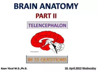
3rd part: Cerebrum • Largest part of brain • Higher thinking takes place • Looks like cauliflower • Creatively controls the environment over animals • Cerebrum divided into halves called Cerebral hemispheres
chapter 4 The cerebrum Largest brain structure Two cerebral hemispheres connected by the corpus callosum. In charge of most sensory, motor, and cognitive processes Surrounded by cerebral cortex, a collection of several thin layers of cells (gray matter)
Cerebrum Cont… • Hemispheres connected by bands of fibers called Corpus Callosum (CORE-pus- ca-LOW-suhm) • Left side controls right side of body and vise versa • However two hemispheres have different talents and tasks, called Lateralization
Cerebral Cortex • Thin layer of dense packed cells, cover Cerebellum • Top of the Brain! • Latin for bark • Produce grey tissue so grey matter • 1/8th of inch thick • But ¾ of cells in human brain
chapter 4 Lobes of the cerebral cortex Occipital lobes (visual cortex) Parietal lobes (somatosensory cortex) Temporal lobes Memory, perception, emotion, and auditory cortex Left lobe: Wernicke’s area Frontal lobes Emotion, planning, creative thinking, and motor cortex Left lobe: Broca’s area
Lobes of the Cortex • 4 distinct regions • Occipital (ahk-SIP-uh-tuhl) lobes • Back of head, visual cortex= vision • Visual signals processed • Damage to visual cortex impair vision, blindness
Parietal Lobes • Somatosensory cortex • Receives information about pressure, pain, touch, temp. • Signals from hands, face • Attention and mental operations
Temporal Lobes • Latin for temples • Sides of brain, just above the ears • Contain audio cortex- processes sounds • Wernicke’s area- involved in language comprehension
Frontal lobes • Front of brains cerebral cortex • Motor cortex, controls 600 muscles in body • Short term memory, higher order thinking, initiative, social judgment • Broca’s Area- speech production
chapter 4 Lobes of the cerebral cortex
Cortex cont… • Stimulate with electrical current to parietal lobes= feel in skin; visual cortex, oct. lobes= flash, light • But some areas stimulated do nothing association cortex, higher mental process • Prefrontal cortex- barely exists in mice, 7 % in dogs, but 29% in humans
chapter 4 Your turn Jenny bumps her head and is suddenly unable to see, although the doctor says there is nothing wrong with her eyes? Which part of her brain did Jenny damage? 1. The amygdala 2. The hippocampus 3. The occipital lobe of the cerebral cortex 4. The parietal lobe of the cerebral cortex
chapter 4 Your turn Jenny bumps her head and is suddenly unable to see, although the doctor says there is nothing wrong with her eyes? Which part of her brain did Jenny damage? 1. The amygdala 2. The hippocampus 3. The occipital lobe of the cerebral cortex 4. The parietal lobe of the cerebral cortex
chapter 4 Phineas Gage Gage was a railroad construction foreman An 1848 explosion forced a steel tamping rod through his head Others said he was “no longer Gage” Lost his job, worked as a sideshow exhibit
Summary Brain part 2 • Limbic system; amygdala, hippo( gage), hypo. • 4 cortexes