Download
1 / 19
531 likes | 1.91k Vues
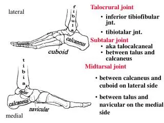
Talocrural joint inferior tibiofibular jnt. tibiotalar jnt. lateral. fibula. calcaneus. Subtalar joint aka talocalcaneal between talus and calcaneus. cuboid. Midtarsal joint between calcaneus and cuboid on lateral side between talus and navicular on the medial side. tibia. talus.

E N D
Talocrural joint • inferior tibiofibular jnt. • tibiotalar jnt. lateral fibula calcaneus • Subtalar joint • aka talocalcaneal • between talus and calcaneus cuboid • Midtarsal joint • between calcaneus and cuboid on lateral side • between talus and navicular on the medial side tibia talus calcaneus navicular medial
Talocrural Joint • Articulation between the tibia and fibula (inferior tibiofibular joint) and between the tibia and the talus (tibiotalar joint). • This joint is responsible for plantar flexion and dorsiflexion and some abduction/adduction. • The axis of rotation is a line between the two malleoli.
Subtalar Joint • Allows pronation/supination and rotation. • The talus articulates with the calcaneus anteriorly, posteriorly and medially. • The axis of rotation runs diagonally from the posterior, lateral, plantar surface to the anterior, medial, dorsal surface. • The orientation of this axis makes pronation/supination triplanar with reference to the cardinal planes.
calcaneus moves on talus talus moves on calcaneus Pronation/Supination Pronation Supination Open Chain calcaneal eversion calcaneal inversion abduction adduction dorsiflexion plantar flexion Closed Chain calcaneal eversion calcaneal inversion talar adduction talar abduction plantar flexion dorsiflexion
Tibial Rotation • The subtalar joint can be likened to the action of a mitered hinge (Inman and Mann, 1973). • The orientation of the subtalar joint axis causes the tibia to internally rotate during pronation and externally rotate during supination. • Thus, the tibia internally rotates with pronation or knee flexion and externally rotates with supination or knee extension. It is important that knee flexion and pronation occur in synchronization (as well as knee extension and supination).
Midtarsal Joint During pronation, the axes of these two joints are parallel, this unlocks the joint and creates a hypermobile foot that can absorb shock. During supination the axes are not parallel and this joint becomes locked allowing efficient transmission of forces. Actually consists of two joints: the calcaneocuboid on the lateral side and the talonavicular on the medial side.
A forefoot valgus exists when the forefoot is everted relative the rearfoot. This is not as common as forefoot varus. A forefoot varus exists when the forefoot is inverted to the rearfoot. This is the most common cause of excessive pronation.
A rearfoot valgus exists when the rearfoot is everted. A rearfoot varus exists when the rearfoot is inverted. This can increase maximum pronation.
Ligaments Lateral side of ankle accounts for 85% of ankle sprains
Arches of the Foot Fascia Plantar surface There are 3 arches in the foot that contribute to support and shock absorption. These arches are maintained by the shape of the tarsal and metatarsal bones, ligaments and plantar fascia.
Arch Types • Feet are often classified according to the height of the medial arch. • Normal • high-arched or pes cavus • flat-footed or pes planus • Arches can also be rigid or flexible. • High-arched, rigid feet make poor shock absorbers. • Flat-footed, flexible arches often allow excessive pronation.
Plantar Flexors Gastrocnemius Soleus NOTE: 1) Soleus lies deep to gastrocnemius 2) Both insert into the calcaneal tendon aka Achilles tendon Posterior View
Assistant Plantar Flexors Note: Their tendons pass posteriorly to the malleoli Flexor Digitorum Longus Flexor Hallucis Longus Tibialis Posterior Peroneus Brevis Peroneus Longus Plantaris Note: insertion is wrong!
Dorsiflexion tibialis anterior extensor hallucis longus (deep to ext. digitorum longus) extensor digitorum longus peroneus tertius (usually very close to extensor digitorum longus and often considered as part of this muscle)
Invertors NOTE: Muscles pass to the medial side of the foot! primary extensor hallucis longus flexor digitorum longus flexor hallucis longus tibialis anterior tibialis posterior
Evertors primary extensor digitorum longus peroneus longus peroneus tertius peroneus brevis
Causes of Excessive Pronation • Q-angle greater than 20 degrees • tibial varus greater than 5 degrees • rearfoot varus greater than 2 degrees • forefoot varus greater than 3 degrees • plantar flexed first ray • weak medial arch • tight gastrocnemius and soleus or a short Achilles tendon
The Problem with Excessive Pronation Excessive or prolonged pronation during the support phase will disrupt the normal tibial-femoral rotation relationship at the knee. The tibia continues to internally rotate with the prolonged pronation while the knee is extending. Knee extension is normally associated with external tibial rotation.