Download
1 / 39
390 likes | 503 Vues
The Role of the Proximal Tail in the Large Steps of Myosin VI. Ron Rock University of Chicago. Myosin II and F-Actin Architecture. Catalytic Domain. Coiled Coil. RLC. ELC. Myosin II: Hexamer of 2 Heavy Chains & 4 Light Chains. F-Actin: Polymer of actin monomers. 36 nm. Pointed End.

E N D
The Role of the Proximal Tail in the Large Steps of Myosin VI Ron Rock University of Chicago
Myosin II and F-Actin Architecture Catalytic Domain Coiled Coil RLC ELC Myosin II: Hexamer of 2 Heavy Chains & 4 Light Chains F-Actin: Polymer of actin monomers 36 nm Pointed End Barbed End
D Step Size and the Lever Arm The light chain binding domain is believed to rotate upon binding to actin Result: Small structural changes in the catalytic domain are amplified
Step Size Correlates to Lever Arm Length for Myosin II • Velocities in gliding filament assays correlate Uyeda et al. PNAS 93 4459 (1996) • Step sizes correlate Warshaw et al. JBC 275 37167 (2000) Ruff et al. Nat. Str. Biol. 8 226 (2001)
A Hand-Over-Hand Model • Myosin V and VI walk… • … in a hand-over-hand manner … • … using two catalytic heads
A Hand-Over-Hand Model D rate-limiting T P
Hand-Over-Hand Motility Matthew L. Walker, Stan A. Burgess, James R. Sellers, Fei Wang, John A. Hammer III, John Trinick & Peter J. Knight. Nature, 405 , 804-807 (2000).
Two Actin Tracks Myosin V Can Cross Actin Filaments
How Does Myosin V Cross Filaments? Flexibility here? Side View Diffusive Search? Top View
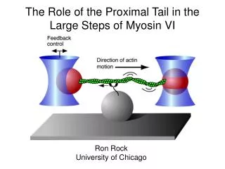
Optical Trap Design Brightfield Trap Steering
Myosin V Stepping in the Trap 480 400 Displacement (nm) 320 240 160 80 5.45 5.5 5.55 Time [s] 0 0 1 2 3 4 5 6 Time (s) 2 mM ATP
Myosin V and VI Takes Large Steps • Mean step is near the actin helical repeat • (VI) Large steps, much larger than expected from lever arm model • (VI) Distribution very broad (30 ± 12 nm) • (VI) Many backsteps (toward barbed end) • (-13 ± 8 nm) VI V
Myosin V and VI Stepping Model Coiled-coil unfolds
The two heads of myosin VI can separate 27 ± 6 nm (SD)
Less than half of the processive stepsize is generated by the working stroke Similar to Myosin V: Veigel et al., Nat. Struct. Bio. 4 59 (2002)
The proximal tail does not act as a rigid lever arm N = 195 11.9 ± 1.2 nm (SE)
The proximal tail is exposed and sensitive to proteolysis by V8 protease Solid arrows indicate 97 kD band. Uncut Myosin VI is 146 kD. Actin:myosin is at 6:1 mol ratio and nucleotides are at 2 mM unless indicated.
The proximal tail allows separation of the heads to produce a large step M6-2hepzip V858 to S888 -> GCN4 19 ± 2 nm (SD)
The proximal tail allows separation of the heads to produce a large step
80 AA => 28.8 nm (contour length) each stiffness k = 0.3 pN/nm (WLC, Lp = 0.9 nm, constant over these ranges) First passage time under zero ext. load (26 nm) is ~6 ms Under 2 pN load, first passage time is 3 s Myosin VI stepping model • Dock proximal tail along the actin filament • Alpha helical proximal tail
Acknowledgments Protein production, EM, kinetics Bhagavathi Ramamurthy Sara Beccafico Carl Morris Clara Franzini-Armstrong H. Lee Sweeney Optical Trapping, proteolysis Alex Dunn Ben Spink Bhadresh Rami Jim Spudich The Helen Hay Whitney Foundation The Burroughs Wellcome Fund
Full-length myosin VI is a monomer A form of motor regulation like Unc104?
ADP Rigor EM of Myosin VI Decorated Actin Shows Evidence of Left Handed Rotation Pointed End Barbed End Wells et al. Nature 401 505 (1999)
F = 1.7 pN 30 nm F = 1.7 pN 30 nm F = 1.7 pN 30 nm Load and the Diffusive Search 50 pN•nm = 200,000x slower 0 pN•nm 25 pN•nm = 400x slower
ADP Release is the Rate Limiting Transition ADP 10 µM ATP ( = Km) k0 = 9 s-1,k1 =17 s-1 2 mM ATP, 400 µM ADP k0 = 6.4 s-1,k1 =161 s-1
Single Fluorophore Detection Nd:YAG 532 nm HeNe 633 nm Ar Ion 488 nm Adapted from Tokunaga BBRC 235 47 (1997)