Ch. 24 Digestive System
Ch. 24 Digestive System. Anatomy. Tooth Structure. • Two main regions – crown and the root • Crown – exposed part of the tooth above the gingiva (gum)

Ch. 24 Digestive System
E N D
Presentation Transcript
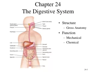
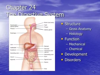
Ch. 24 Digestive System Anatomy
Tooth Structure • • Two main regions – crown and the root • • Crown – exposed part of the tooth above the gingiva (gum) • • Enamel – acelluar, brittle material composed of calcium salts and hydroxyapatite crystals is the hardest substance in the body • • Encapsules the crown of the tooth • • Root – portion of the tooth embedded in the jawbone • • Neck – constriction where the crown and root come together
• Cementum – calcified connective tissue • • Covers the root • • Attaches it to the periodontal ligament • • Periodontal ligament • • Anchors the tooth in the alveolus of the jaw • • Forms the fibrous joint called a gomaphosis • • Gingival sulcus – depression where the gingival borders the tooth • • Dentin – bonelike material deep to the enamel cap that forms the bulk of the tooth • • Pulp cavity – cavity surrounded by dentin that contains pulp • • Pulp – connective tissue, blood vessels, and nerves
• Root canal – portion of the pulp cavity that extends into the root • • Apical foramen – proximal opening to the root canal • • Odontoblasts – secrete and maintain dentin throughout life
Tooth and Gum Disease • • Dental caries – gradual demineralization of enamel and dentin by bacterial action • • Dental plaque, a film of sugar, bacteria, and mouth debris, adheres to teeth • • Acid produced by the bacteria in the plaque dissolves calcium salts • • Without these salts, organic matter is digested by proteolytic enzymes • • Daily flossing and brushing help prevent caries by removing forming plaque
Tooth and Gum Disease: Periodontitis • • Gingivitis – as plaque accumulates, it calcifies and forms calculus, or tartar • • Accumulation of calculus: • • Disrupts the seal between the gingivae and the teeth • • Puts the gums at risk for infection • • Periodontitis – serious gum disease resulting from an immune response
Meth Mouth • When meth is ingested, it causes the user's blood vessels to shrink, limiting the steady blood supply that the mouth needs in order to stay healthy. With repeated shrinking, these vessels die and the oral tissues decay. Similarly, meth use leads to "dry mouth" (xerostomia), and without enough saliva to neutralize the mouth's harsh acids, those acids eat away at the tooth and gums, causing weak spots that are susceptible to cavities. The cavities are then exacerbated by behavior common in users on a meth high: a strong desire for sugary foods and drinks, compulsive tooth grinding, and the general neglect of regular brushing and flossing.
Faces of Meth • http://facesofmeth.us/main.htm
Pharynx • • From the mouth, the oro- and laryngopharynx allow passage of: • • Food and fluids to the esophagus • • Air to the trachea
Esophagus • • Muscular tube going from the laryngopharynx to the stomach • • Travels through the mediastinum and pierces the diaphragm • • Joins the stomach at the cardiac orifice
Homeostatic Imbalance • • Heartburn (gastroesophageal reflux disease or GERD) – burning, radiating substernal pain caused by acidic gastric juice regurgitated into the esophagus • • Caused by excess eating or drinking, and conditions that force abdominal contents superiorly (e.g., extreme obesity, pregnancy, and running) • • Hiatus hernia – structural abnormality in which the superior part of the stomach protrudes slightly above the diaphragm • • Prolonged episodes can lead to esophagitis, ulcers, and cancer
Digestive Processes in the Mouth • • Food is ingested • • Mechanical digestion begins (chewing) • • Propulsion is initiated by swallowing • • Salivary amylase begins chemical breakdown of starch • • The pharynx and esophagus serve as conduits to pass food from the mouth to the stomach
Stomach • • Chemical breakdown of proteins begins and food is converted to chyme • • Cardiac region – surrounds the cardiac orifice • • Fundus – dome-shaped region beneath the diaphragm • • Body – midportion of the stomach • • Pyloric region – made up of the antrum and canal which terminates at the pylorus • • The pylorus is continuous with the duodenum through the pyloric sphincter • • Greater curvature – entire extent of the convex lateral surface • • Lesser curvature – concave medial surface • • Lesser omentum – runs from the liver to the lesser curvature • • Greater omentum – drapes inferiorly from the greater curvature to the small intestine • • Nerve supply – sympathetic and parasympathetic fibers of the autonomic nervous system • • Blood supply – celiac trunk, and corresponding veins (part of the hepatic portal system)
Microscopic Anatomy of the Stomach • •Muscularis – has an additional oblique layer that • • Allows the stomach to churn, mix and pummel food physically • • Breaks down food into smaller fragments • • Epithelial lining is composed of: • • Goblet cells that produce a coat of alkaline mucus • • Gastric pits containing gastric glands that secrete: • • Gastric juice • • Mucus • • Gastrin
Glands of the Stomach Fundus and Body • • Gastric glands of the fundus and body have a variety of secretory cells • • Mucous neck cells – secrete acid mucus • • Parietal (oxyntic) cells – secrete HCl and intrinsic factor • • Chief (zymogenic) cells – produce pepsinogen • • Pepsinogen is activated to pepsin by: • • HCl in the stomach • • Pepsin itself by a positive feedback mechanism • • Enteroendocrine cells – secrete gastrin, histamine, endorphins, serotonin, cholecystokinin (CCK), and somatostatin into the lamina propria
Stomach Lining • • The stomach is exposed to the harshest conditions in the digestive tract • • To keep from digesting itself, the stomach has a mucosal barrier with: • • A thick coat of bicarbonate-rich mucus on the stomach wall • • Epithelial cells that are joined by tight junctions • • Gastric glands that have cells impermeable to HCl • • Damaged epithelial cells are quickly replaced
Digestion in the Stomach • • The stomach: • • Holds ingested food • • Degrades it both physically and chemically • • Delivers chyme to the small intestine • • Enzymatically digests proteins with pepsin • • Secretes intrinsic factor required for absorption of vitamin B12
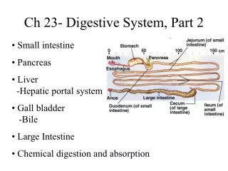
Small Intestine: Gross Anatomy • • Runs from pyloric sphincter to the ileocecal valve • • Has three subdivisions: duodenum, jejunum, and ileum • • The bile duct and main pancreatic duct: • • Join the duodenum at the hepatopancreaticampulla • • Are controlled by the sphincter of Oddi • • The jejunum extends from the duodenum to the ileum • • The ileum joins the large intestine at the ileocecal valve
Liver • • The largest gland in the body • • Superficially has four lobes – right, left, caudate, and quadrate • • The falciform ligament: • • Separates the right and left lobes anteriorly • • Suspends the liver from the diaphragm and anterior abdominal wall • • The ligamentumteres: • • Is a remnant of the fetal umbilical vein • • Runs along the free edge of the falciform ligament
Liver: Associated Structures • • The lesser omentum anchors the liver to the stomach • • The hepatic blood vessels enter the liver at the portahepatis • • The gallbladder rests in a recess on the inferior surface of the right lobe • • Bile leaves the liver via • • Bile ducts which fuse into the common hepatic duct • • The common hepatic duct fuses with the cystic duct • • These two ducts form the bile duct
Homeostatic Imbalance • • Hepatitis – inflammation of the liver often due to viral infection • • Viruses causing hepatitis are catalogued has HVA through HVF • • HVA and HVE are transmitted enterically and cause self-limiting infections • • Hepatitis B is transmitted via blood transfusions, contaminated needles, and sexual contact, and increases the risk of liver cancer • • Hepatitis C produces chronic liver infection • • Nonviral hepatitis is caused by drug toxicity and wild mushroom poisoning • • Cirrhosis – diffuse and progressive chronic inflammation of the liver • • Typically results from chronic alcoholism or severe chronic hepatitis • • The liver becomes fatty and fibrous and its activity is depressed • • Scar tissue obstructs blood flow in the hepatic portal system causing portal hypertension
The Gallbladder • • Thin-walled, green muscular sac on the ventral surface of the liver • • Stores and concentrates bile by absorbing its water and ions • • Releases bile via the cystic duct which flows into the bile duct
Homeostatic Imbalance • • Gallstones – crystallization of cholesterol which can obstruct the flow of bile • • Current treatments include: dissolving the crystals with drugs, pulverizing them with ultrasound, vaporizing them with lasers, and surgical removal of the gallbladder • • Obstructive jaundice – yellowish skin caused by bile pigments deposited in the skin • • Due to blocked bile ducts
Pancreas • • Location • • Lies deep to the greater curvature of the stomach • • The head is encircled by the duodenum and the tail abuts the spleen • • Exocrine function • • Secretes pancreatic juice which breaks down all categories of foodstuff • • Acini (clusters of secretory cells) contain zymogen granules with digestive enzymes • • The pancreas also has an endocrine function – release of insulin and glucagon
Large Intestine • • Is subdivided into the cecum, appendix, colon, rectum, and anal canal • • The saclike cecum: • • Lies below the ileocecal valve in the right iliac fossa • • Contains a wormlike vermiform appendix
Homeostatic Imbalance • • Appendicitis – inflammation of the appendix resulting from blockage that traps infectious bacteria in its lumen • • If the appendix ruptures, feces containing bacteria spray over the abdominal contents causing peritonitis • • Treatment is surgical removal of the appendix
Colon • • Has distinct regions: ascending colon, hepatic flexure, transverse colon, splenic flexure, descending colon, and sigmoid colon • • The transverse and sigmoid portions are anchored via mesenteries called mesocolons • • The sigmoid colon joins the rectum • • The anal canal, the last segment of the large intestine, opens to the exterior at the anus
Valves and Sphincters of the Rectum and Anus • • Three valves of the rectum stop feces from being passed with gas • • The anus has two sphincters: • • Internal anal sphincter composed of smooth muscle • • External anal sphincter composed of skeletal muscle • • These sphincters are closed except during defecation
Functions of the Large Intestine • • Other than digestion of enteric bacteria, no further digestion takes place • • Vitamins, water, and electrolytes are reclaimed • • Its major function is propulsion of fecal material toward the anus • • Though essential for comfort, the colon is not essential for life
Homeostatic Imbalance • • Diverticulosis – small herniation (diverticula) of the mucosa of the colon walls caused by lack of bulk in the colon • • Most common in the sigmoid colon in people over 70 • • Diverticulitis – inflamed diverticula that can be life threatening if the diverticula rupture
Defecation • • Distension of rectal walls caused by feces • • Stimulates contraction of the rectal walls • • Relaxes the internal anal sphincter • • Voluntary signals stimulate relaxation of the external anal sphincter and defecation occurs
Homeostatic Imbalance • • Diarrhea – watery stool resulting from any condition that rushes food residue through the large intestine too quickly • • This causes insufficient time for water absorption • • Prolonged diarrhea may result in dehydration and electrolyte imbalance • • Constipation – hard stool that is difficult to pass resulting from residues staying in the intestine too long • • May result from lack of fiber in the diet
Food Poisoning: Salmonella • Food Poisoning: Salmonella • • Salmonella is spread by: • • Contaminated eggs and egg products • • Infected food handlers with feces-contaminated hands • • Salmonella can cause: • • Bacteremia 4 to 7 days after infection • • Endocarditis, thrombi, bone infections, arthritis, and meningitis • • Diagnosis is by positive stool samples • • Salmonellosis is treated symptomatically
Homeostatic Imbalance • • Cleft palate – palatine bones, palatine process of the maxillae (or both) fail to fuse • • Tracheoesophageal fistula – opening between the esophagus and trachea • • Cystic fibrosis – impairs pancreatic activity
Cancer • • Stomach and colon cancers rarely have early signs or symptoms • • Metastasized colon cancers frequently cause secondary liver cancer • • Prevention is by regular dental and medical examinations • • Colon cancer is the 2nd largest cause of cancer deaths in males (lung cancer is 1st) • • Forms from benign mucosal tumors called polyps whose formation increases with age • • Regular colon examination should be done for all those over 50