Small Intestine and Pancreas: Digestive System Essentials
640 likes | 673 Vues
Explore the crucial roles of the small intestine and pancreas in chemical digestion and nutrient absorption. Learn about the unique features, enzymes, and functions of these digestive organs. Understand the hepatic portal system and liver's metabolic functions.

Small Intestine and Pancreas: Digestive System Essentials
E N D
Presentation Transcript
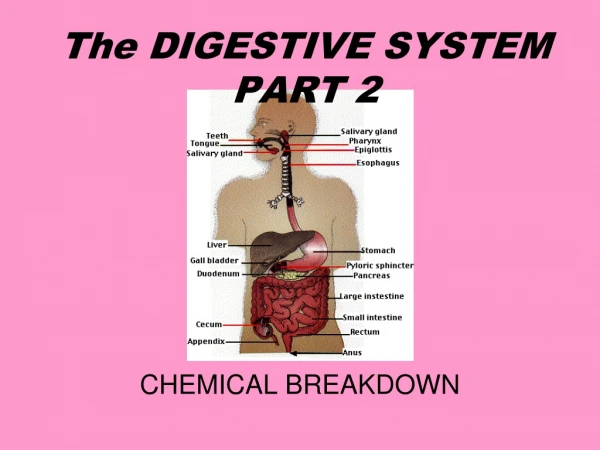
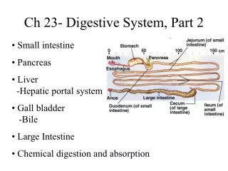
Small intestine • Pancreas • Liver -Hepatic portal system • Gall bladder -Bile • Large Intestine • Chemical digestion and absorption Ch 23- Digestive System, Part 2
Small Intestine • Major function- responsible for 90% of absorption and also important for digestion • Divided into three regions • Duodenum- ‘mixing bowl’ • Jejunum- chemical digestion/absorption • Ileum- protection from large intestine
Layers of the small intestine Plica Unique to the small intestine is the presence of plica (plicae circulares). Plica are permanent folds of the mucosa and submucosa.
One plica One villus On the plica, you will see villi--Present only in small intestine -Crypts spaced between them -Do not confuse with ‘mircovilli’
Plica and villi Each plica supports a forest of villi. Each villus is covered with simple columnar epithelium. These epithelial cells have microvilli. Phew!
Villus and enzymes of the brush border cells microvilli Brush border cell • The lamina propria of each villus contains a lacteal, nerves and blood vessels
Regional Features of the Small Intestine • Duodenum- site of chyme entry, where it is blasted with HCO3- (and other things) • Along duodenum, pH of chyme changes from 1-2 to 7-8 • Mucus glands of mucosa • Also, Brunner’s glands (submucosal glands) • Jejunum- first half has numerous plicae and villi, but they decrease in number en route to ileum • Brush border enzymes • Ileum - lack plicae, contain masses of lymphoid tissue..why??
Duodenum Villi Crypts Plica Brunner’s Glands (submucosal glands)
Ileum Goblet Cells Crypts Peyer’s Patches
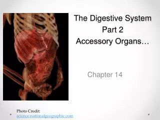
What organs secrete into the small intestine? Liver Gall bladder
Anatomy of the pancreas -An accessory structure of the small intestine
Pancreas • Where have you seen the pancreas before? • Primarily an exocrine gland (99% of cells) • Composed of a series of ductules that are surrounded by secretory cells, acinar cells
Acinar Cells • Cuboidal epithelial cells that form pancreatic acini • Pancreatic juice produced by acinar cells • Alkaline mixture of enzymes, water, and ions • Over 1200 ml of ‘juice’ produced each day! • Travels in pancreatic duct, joins the common bile duct
Enzymes produced by the pancreas • Pancreatic alpha-amylase • Pancreatic lipase • Nucleases • Four inactive proteolytic enzymes • Not activated until they reach the small intestine
Proteolytic Enzymes • Inactive trypsinogen converted to active trypsin by enteropeptidase (enterokinase) • Trypsin then converts other proenzymes into their active forms • Chymotrypsin • Carboxypeptidase
Inactive proteases released from acinar cells 1 2 3 trypsin 6 TRYPSIN trypsin trypsinogen 5 Inactive protease active protease 4 enteropeptidase • Trypsin is now active, and will activate other proteases: • Chymotrypsin • Carboxypeptidase Brush border cells
Liver • Largest visceral organ, 3 lb • Surrounded by fibrous capsule • Has numerous lobes, with numerous lobules • Cells are called hepatocyes • Receives ~25% of total cardiac output!! • Blood flow to and from the liver is unique
Hepatic Portal System • Liver receives 1/3 of blood via hepatic artery (not shown) • 2/3 from hepatic portal vein • This major vein drains capillaries from: esophagus, stomach, small and large intestine and THEN heads to liver • Venous blood from liver dumps directly into inferior vena cava Any guesses as to a major function of the liver?
Hepatic Portal System -The portal venous blood contains all of the products of digestion absorbed from the GI tract. -Thus all nutrients are processed/viewed in the liver (by hepatocytes) before being released back into the central veins.
Kupffer RBCs Liver • Each lobe divided into lobules • Cells of the liver are hepatocytes • Arranged as a single layer of cells, stacked • Divided by sinusoids • Blood flows through sinusoids, past Kupffer cells (resident macrophage) bacteria
Liver lobules Blood is flowing towards center Hepatocytes arranged into ‘walls’. Walls radiate outward from a central point. The central area contains a single, central vein.
1 mm Liver lobules- Portal Triads Central Vein Portal Triad
Major functions of the liver • Metabolic regulation • Hematological regulation • Bile production (its only truedigestive function)
Metabolic Regulation • Hepatocytes regulate the composition of the blood and blood nutrients • They do this by selectively secreting and absorbing molecules from the blood • What molecules? Carbs, lipids, amino acids, waste products, vitamins, minerals and drugs Portal triad
Metabolic regulation • Hepatocytes adjust the contents of the blood by selective absorption and secretion • Remove/store excess nutrients • Correct deficiencies • Hepatocytes are able to take in and/or release large molecules with ease, partly because the nearby capillaries are so leaky! hepatocytes A big ol’ leaky capillary, a sinusoidal capillary
What role does the liver play in amino acid metabolism? • Removes excess amino acids from the bloodstream or releases stored amino acids that are deficient • Besides maintenance/growth, what might these amino acids be used for? Alanine, valine, leucine, proline, etc, etc.
What role does the liver play in lipid metabolism? • Regulates circulating levels of triglycerides, fatty acids, and cholesterol • If levels decline, lipid stores are broken down and released • ALSO, we will return to Bile in 3 minutes, an important player in lipid breakdown
What role does the liver play in carbohydrate metabolism? • Blood glucose levels are kept at or near 90 mg/dl • If blood glucose levels rise, glucose is stored in liveras ______? • If glucose levels drop, glycogen reserves in the liver are broken down and glucose is released into blood
Other metabolic functions of the liver • Removal of waste products • Ammonia is converted to urea • Vitamin storage • The fat soluble ones! • Mineral storage • Drug inactivation
Kupffer RBCs Hematological regulation • Synthesis of plasma proteins (think albumins, etc.) • Removal of hormones (like a giant sponge, the liver takes NE, epi, thyroid hormones, steroid hormones, etc. ‘out of the game’) • Removal of antibodies • Removal (or storage) of toxins • Phagocytosis (Kupffer cells chomp up RBCs and are APCs!) • Production of bile (1 liter/day)
Hepatocytes produce bile • Bile is made and then secreted into channels called bile canaliculi • Canaliculi merge to join the bile ducts at each of the triads triad
What is bile? • Bile is an alkaline solution containing: water, electrolytes, bilirubin, phospholipids, triglycerides, cholesterol, and lipids known as bile salts • Function of bile (bile salts):emulsify fats for digestion • Bile salts are not enzymes, they merely help to bust up large fat droplets into smaller fat droplets. • Why is this important?
Gall Bladder • Major functions: • Bile storage • Bile concentration, modification Gall bladder receives bile from liver via the cystic duct
What regulates release of bile? • When chyme enters the small intestine, CCK is released • CCK relaxes sphincter (sphincter of Oddi) and triggers muscle contractions that squeeze the gall bladder and send bile out, into duodenum • How clever that CCK secretion increases when chyme is high in fats! • Interesting note: CCK also slows stomach movement (release of food to duodenum) • What does this tell you about a high fat meal?
Review of Digestion at Small Intestine • Chyme enters duodenum from stomach • Gets blasted with HCO3-, enzymes and bile • Which enzymes? Where from? Are all enzymes active upon arrival? Which ones are? Which ones are not? • Segmentation provides mechanical digestion • Bile salts surround fats, increasing lipase effectiveness • ABSORBPTION of nutrients!! • Once most absorption done, two waves of peristalsis push left-overs into large intestines. Each wave takes ~2 hours. Animation-Recap
The last part..large intestine • General functions • Reabsorption of water • Absorption of vitamins made by bacteria • Compaction and storage of fecal material
Anatomy of the large intestine The large intestine is comprised of sections: • Cecum and appendix • Colon (ascending, transverse, descending, sigmoid) • Rectum, leads to anal canal and anal canal Note-Large diameter Thin walls
Histology of the Large Intestine • Colon- Simple columnar with goblet cells • Rectum- the anal canal is stratified squamous • So, as you move from colon to out: epithelium changes from columnar (with goblet cells) stratified squamous keratinized stratified squamous. • Mucosa does not produce enzymes • No villi Site of-Vitamins, bile, and some H2O absorption
A word about bacterial flora Most bacteria are DOA (dead on arrival) at the large intestine, but some survive and comprise the bacterial flora Over 700 species! Functions?
Chemical Digestion and Absorption • Virtually all nutrients from the diet are absorbed into blood (or lymph vessels) across the mucosa of the small intestine. • Much of this absorption happens via co-transport or diffusion across the apical membrane of intestinal epithelial cells (the brush border cells), aka eneterocytes
Chemical digestion and absorption Part 1 & 2 First steps of mechanical and chemical digestion Part 3 Final steps of chemical digestion and absorption
Hydrolysis • Hydrolysis is breaking of a bond ‘with water’ or the addition of water • Digestive enzymes hydrolyze one specific substrate A-B-C-D + H2O A-B-H + OH-C-D ABCDase
Absorption • Carbohydrates- simple, hexose sugars will be absorbed • Proteins- di or tripeptides are typically absorbed, also amino acids • Fats- Fatty acids and monoglycerides are absorbed (and then converted immediately back to triglycerides)
Carbohydrates • Sucrase catalyzes the cleavage of the bond between (glucose-fructose) in sucrose • Lactase catalyzes the cleavage of the bond between (glucose-galactose) in lactose • Maltase catalyzes the cleavage of the bond between (glucose-glucose) in maltose
Where do we find these three enzymes? • Enzymes are transmembrane (or at least membrane-associated) proteins • Function at the microvilli of enterocytes (brush border cells) • Apical surface Maltase Lactase Sucrase brush border cells
Disaccharides in intestinal lumen interact with brush border enzymes, and the enzymatic products are absorbed Hexose sugars Hexose sugars Hexose sugars Maltose Sucrose Lactose Maltase Sucrase Lactase brush border cells
When brush border epithelial cells fail to make lactase…. Lactose is not digested or absorbed. Bacteria ferment lactose Gas, diarrhea, etc. Sucrase Maltase Lactase