Digestive System Part 3
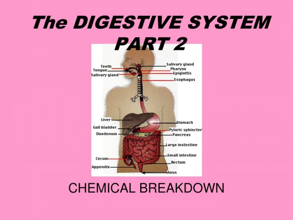
Digestive System Part 3. Small Intestine. Duodenum: smallest section Stomach empties here Jejunum: middle section Ileum: final section Joins large intestine at ileocecal sphincter. Small Intestine. 21 feet of small intestine with added structures to increase surface area

Digestive System Part 3
E N D
Presentation Transcript
Digestive System Part 3
Small Intestine • Duodenum: smallest section • Stomach empties here • Jejunum: middle section • Ileum: final section • Joins large intestine at ileocecalsphincter
Small Intestine • 21 feet of small intestine with added structures to increase surface area • Almost all absorption occurs here • Circular folds: folds in the mucosa to splash chyme and enhance absorption • Villi: fingerlike projections that contain capillaries, arteries, veins, all to move absorbed substances quickly
Small Intestine • Microvilli: smaller projections on the villi to further increase surface area and absorption
Small Intestine • Intestinal glands: in mucosa that secrete intestinal juice • Clear, yellowish liquid quickly reabsorbed • pH 7.6 with water and mucous • Enzymes for Chemical Digestion: • Maltase, sucrose, lactase: disac. To monosa. • Peptidases: peptides to amino acids • Ribonuclease, dioxyribonuclease: nucleic acids
Small Intestine • Mechanical Digestion: • Segmentation: Concentration of chyme and juice that sloshes between areas of contraction of the muscularis • Perstalsis: moves the chyme steadily through intestines
Absorption in Small Intestine • Passage of digested substances and nutrients from the lumen to into the blood or lymph • 90% of all absorption takes place here • Substances absorbed by diffusion, osmosis, and active transport
Absorption in Small Intestine • Carbohydrates absorbed as monosaccharides • Proteins absorbed as amino acids • Lipids absorbed as monoglycerides and fatty acids • Water —9 liters enter daily and 8 are reabsorbed
Large Intestine • Main functions: • Completion of absorption • Manufacture certain vitamins • Formation of feces • Elimination of feces
Large Intestine • Cecum: pouch after ileocecal sphincter • Colon: ascending, transverse, descending, sigmoid • Surface area increased by pouch-like divisions: haustra • Rectum: last part, stores waste • Last inch is anal canal • Anus: opening to exterior • Two sphincters: internal anal sphincter (involuntary), external anal sphincter (voluntary)
Chemical Digestion of Lrg. Intestine • No enzymes • Bacteria ferment remaining carbohydrates • Release hydrogen, carbon dioxide, and methane • Vitamins K and some B’s are made and absorbed
Mechanical Digestion of Lrg. Intestine • Haustral churning: walls contract when haustra fill to a certain level • Peristalsis: slower than other areas • Mass peristalsis: strong muscular wave that pushes waste into the rectum
Absorption & Feces Formation • Chyme in the colon for 3-10 hrs. becomes rather solid: feces • Water, epithelial cells from mucosa, bacteria, undigested food • All but 100mL of the 1L of water is absorbed
Defecation • Emptying of the rectum • Diarrhea: not enough water is absorbed because chyme travels too quickly through intestine • Can cause dehydration • Constipation: feces remains in colon too long, almost all the water is absorbed
Digestive System Attachments • Peritoneum & serosa: secrete slippery fluid to glide organs over each other • Outermost layer of GI tract • Visceral peritoneum: covers some organs in abdominal cavity • Parietal peritoneum: the walls of the abdominal cavity
Digestive System Attachments • Mesentery: binds small intestine to posterior abdominal wall • Mesacolon: binds large intestine to posterior abdominal wall • Falciform ligament: attaches liver to the anterior abdominal wall and diaphragm
Digestive Disorders • Ulcers: lesions in a membrane • Peptic —from gastric juice • Gastric —in stomach • Esophageal —in esophagus • Duodenal —in small intestine • Appendicitis: inflammation of appendix
Digestive Disorders • Cirrhosis: scarred liver due to chronic inflammation • Colitis: inflammation of colon and rectum • Hernia: protrusion of an organ through a membrane or cavity wall