Download
1 / 45
450 likes | 647 Vues
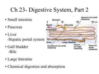
Lecture DIGESTIVE SYSTEM part 2. INTESTINE LIVER PANCREAS. Department of histology, cytology and embryology KhNMU. Small intestine. Functions: digestion – liver, pancreas, enterocytes absorption – enterocytes STRUCTURE: 4 membranes. Intestinal lining:.

E N D
LectureDIGESTIVE SYSTEM part 2 INTESTINE LIVER PANCREAS Department of histology, cytology and embryology KhNMU
Small intestine • Functions: • digestion – liver, pancreas, enterocytes • absorption – enterocytes • STRUCTURE: 4 membranes
Intestinal lining: 1) plicae circularis: mucosa + submucosa 2) villi -consist of: epithelium, lamina propria, mm 3) crypts: invaginations of epithelium in the lamina propria simple columnar epithelium: absorptive enterocytes, goblet, endocrine, Paneth cells and stem cells fenestrated capillaries and central lacteal
Duodenum Ileum lacteal Villus serosa
a Epithelium(villus) bb • high mag. 1). Enterocytes = Columnar absorptive cells (a) have microvilli =brush border (bb) for absorption of digested food. Membrane and luminal digestion 2). Mucus-secreting goblet cells (gc) produce a protective mucus. Lymphocytes and plasma cells (L) are numerous in the lamina propria of the villus. gc L
Epithelium (crypt) • high mag At the bottom of intestinal glands (crypts) are 3). the granule-containing Paneth cells (p). Lysozyme. Goblet (g) and absorptive (a) cells. p g a
3. Paneth cells, 4. enteroendocrine cells (CCK, secretin, GIP), 5.undifferentiated cells=stem, at the bottom of crypt 4 5 3
ivs Villus,lacteal lp l v sm The lamina propria (lp)contains blood-filledcapillariesc, and lacteal –lymphatic capillaryl, smooth muscle cells (sm),(krok – villus shortens); GALT ivs c gc GALT low
v Duodenum low • Mm – 2 layers of sm.m. • Submucosa is usual. Only in the duodenum it is filled with Brunner’s mucous glands (bg), around which - 2 layers of smooth muscle of the muscularis externa (me)surrounded by the serosa(s). m lp ig v mm sm v m me sm med s bg
Duodenum c villi • low & med mag. Ducts (d) from Brunner’s glands (Bg) (s) pass through the muscularis mucosa (mm) to empty their alkaline mucus in or between the crypts (c). d intestinal glands mm mm Bg low s med
Duodenum m Brunner’s glands • nerve supply- submucosal (s) and myenteric (m) nerve plexuses. high s med crypts submucosa muscularis externa muscul. mucosa high
Jejunum v a ig • low & med. mag. The jejunum is like the duodenum & ileum but has no submucosal glands and Peyer’s patches. Contains bigger amount of goblet cells g ig P med low
Ileum cr. v muscul. mucosa • low & med. mag. The major distinguishing feature is the aggregated lymphatic nodules (ln) called Peyer’s patchesin the mucosa or submucosa v sm sm ln med low ln
Duodenum Ileum lacteal Villus COMPARE !
Large intestine (bowel) - general • Reabsorption of water, electrolytes, cellulous • Elimination of wastes • Inner lining - permanent internalfoldsof its mucosa & submucosa called plicae circulares and crypts. • Its mucosa lacks of villi. • The submucosa is usual • Circular & longitudinal smooth muscle form the muscularis externa. The inner circular layer is uniform but the outer longitudinal layer has 3 thicker bands, the taenia coli. • where the colon faces the abdominal cavity there is a serosa.
cr Colon • low & med mag. Plicae circulares (pc) . Unlike the small intestine there are no mucosal villi. There are straight intestinal glands – crypts (cr) composed mainly of 1. Goblet cells – the most numerous . 2. Columnar absorptive cells 3. Enteroendocrine cells 4. Undifferentiated cells muscularis mucosa pc med submucosa low musc. ext.
Large intestineCOMPARE ! • Appendix
ss g Anal canal L • low & high mag. Anal columns Brunched tubular glands (g) Stratified squmous epithelium (ss) v ig mm v a v low gc high
Liver & Gall Bladder Liver has specific location – on the way of absorbed material, that is why has very original vasculature and functions
Functions: • Bile synthesis and secretion (emulsification) • Excretion of bilirubin • Protein synthesis • Gluconeogenesis • Storage • Detoxification • Protective • Hemopoietic organ • Endocrine
Liver lobule is hexagonal in shape ① at its center - central vein ② hepatic plates(cords of cells - hepatocytes)locate radially ③ hepatic sinusoids locate between plates
Connective tissue poor develops Central veins (cv) drainto a sublobular vein (sv). The portal triads (pt) locate at lobule coners. cv sv pt
Hepatic plate(cord)is one or two cells thick • Inside the plate between cells the bile canaliculi locate Bile canaliculus hepatocyte sinusoid Kupffer cell The bile canaliculus wall is made up of hepatocytes
Hepatocytes: Polyhedral 6 surfaces Vascular and bile surfaces a. RER b. SER c. Golgi complex d. Mitochondria e. Inclusions • Jaundice • ①High regeneration
EM of Liver • Liver • Two hepatocytes (h) are seen. • The bile canaliculus form a network that eventually empty into bile duct at the periphery of the lobule. • Ultimately ducts will empty into the gall bladder. h h b
Hepatic sinusoid Lined by endothelium Hepatic macrophages (Kupffer cell) • Space of Disse • sinusoid • Kupffer cell
e d • high mag. Liver had been injected with carbon particles Kupffer cells containing carbon (red arrows) Sinusoids (s) A flattened endothelial cell (e) Space of Disse (d) between it and the hepatocytes s
Blood circulation of the liver • Hepatic artery interlobular artery • Portal vein interlobular vein • hepatic sinusoid • central vein • cv • sublobular vein • hepatic vein • The way of bile draining s d cv v a
cv • Liver, injected with red gelatin to demonstrate the abundant vasculature. central vein (cv), the portal triad (pt) hepatic sinusoids (hs). hs pt
Pancreas. Mixed gland • Functions: • Exocrine • Trypsinogen, pepsinogen, peptidase • Amylase • Lipase • Deoxyribonuclease, ribonuclease • Endocrine
Pancreas – compound acinar serous gland low lobule • Exocrine – 98-99% • Endocrine – 1-2% • Capsule • Septa • Lobules • Interlobular duct • Serous secretory units lobule id med
Septa (c.t.) • Lobules • Interlobular duct • Serous secretory units
Pancreas. Exocrine secretory units • Zymogen granules • Intercalated duct is intercalated into the s.unit = centroacinar cells
Exocrine part Structural features of the acinus: ① Purely serous. ② the presence of centroacinar cells in the center of the acinus Intercalated duct Serous cells Centroacinar cells
Pancreas. Endocrine • Islets of Langerhans, - low & med. mag. Scattered among exocrine secretory units spherical collections of light-staining cords of endocrine cells . low, H&E med, trichrome
Pancreas. Endocrine • islets of Langerhans: • 1. B cell • 2. A cell • 3. D cell • 4. minor cells: PP, D1, EC,
Islets of Langerhans • B - insulin blood glucose 70% • A – glucagon blood glucose 15-20% • D – somatostatin insulin 5-10% glucagon • PP – PP, • D1 – VIP, • EC – secretin, motilin