Lecture on Digestive System
190 likes | 217 Vues
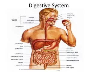
Lecture on Digestive System. www.AssignmentPoint.com. Objectives: Discuss the general functions and anatomy of the digestive tract Describe the individual organs of the system, including a discussion of the gross and microscopic anatomy. Digestive System. consists of:.

Lecture on Digestive System
E N D
Presentation Transcript
Lecture on Digestive System www.AssignmentPoint.com
Objectives: Discuss the general functions and anatomy of the digestive tract Describe the individual organsof the system, including a discussion of the gross and microscopic anatomy.
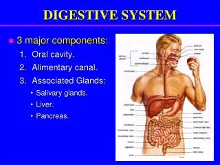
Digestive System consists of: Muscular, hollow tube (= “digestive tract”) + Various accessory organs
Function The function of the system as a whole is processing food in such a way that high energy molecules can be absorbed and residues eliminated. Individual parts function in: • ingestion • mechanical digestion • chemical and enzymatic digestion • secretion • absorption • compaction • excretion and elimination
Histological Organization Tube made up of four layers. Modifications along its length as needed. 1 2 Muscularis externa 3 4
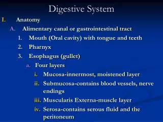
The 4 Layers of the Gut Fig 25.2 1) Mucosa Epithelium – usually simple columnar with goblets; may be stratified squamous if protection needed Lamina propria - connective tissue deep to epithelium Muscularis mucosae -produces folds - plicae (small intestine) or rugae (stomach) 2) Submucosa – made up of loose connective tissue contains submucosal plexus and blood vessels 3) Muscularis externa – smooth muscle, usually two layers (controlled by the myenteric plexus ) - outer layer: longitudinal inner layer: circular 4) Serosa visceral layer of mesentery or adventitia depending on location
Membranes Peritoneum - generic serous membrane in abdominal cavity Mesenteries - double sheets of peritoneum, surrounding and suspending portions of the digestive organs • Greater omentum - "fatty apron", hangs anteriorly from stomach, double layer encloses fat • Lesser omentum - between stomach and liver • Mesentery proper - suspends and wraps the small intestine • Mesocolon - suspends and wraps the colon, parts are i. transverse mesocolon ii. sigmoid mesocolon Fig. 25.4
Oral Cavity • Also called buccal cavity - lined with oral mucosa (type of epithelium ?) • Hard and soft palates - form roof of mouth • Tongue - skeletal muscle • Salivary glands - three pairs • Teeth
Three pairs of Salivary Glands 1-1.5 l / day for digestion (?) lubrication (swallowing) moistening (tasting) • Parotid – lateral side of face, anterior to ear, drain by parotid duct to vestibule near 2nd upper molar • mumps • Submandibular – medial surface of mandible – drain near lingual frenulum drain posterior to lower molars • Sublingual – in floor of mouth - drain near frenulum
Structure of Teeth Crown - exposed surface of tooth Neck - boundary between root and crown Enamel - outer surface Dentin – bone-like, but noncellular Pulp cavity - hollow with blood vessels and nerves Root canal - canal length of root gingival sulcus - where gum and tooth meet Fig 25.7
Types and Numbers of Teeth Dental succession Deciduous (baby, milk) teeth - 20, replaced by Permanent teeth - 32 teeth
Gross Anatomy of the Stomach Lesser curvature Greater curvature Cardia - end under the heart Fundus - bulge above the esophageal opening Body - largest region Pylorus - J curve, inferior end, terminates in Cardiac and Pyloric sphincters (importance?) Rugae – highly extendable interior folds Figs 25-10/11
Histology of Stomach Fig 25.13 Type of epithelium lining stomach? Gastric pits – shallow pits, external half rapidly reproduces for replacement Gastric glands – deep in lamina propria, 3 types of cells • Parietal cells (produce HCl and intrinsic factor) • Chief cells (produce pepsinogen) • Enteroendocrine cells – G cells (several hormones including gastrin which stimulates both parietal and chief cells)
Regions of Small Intestine SI is longest part of dig. tube • Duodenum(short, 12 inches) • fixed shape & position • Mixing bowl for chyme & ? • Jejunum(2.5 m long) • Most of digestion • Ileum (longest at 3.5 m) • Most of absorption, ends in • Ileocecal valve– slit valve into large intestine (colon)
Structure of Small Intestinal Wall Fig 25.15 Plicae circulares – circular pleats around the interior of the small intestine Villi – minute finger-like projections, contain capillaries & lacteals Microvilli – sub-microscopic size, projections on single cells Function of all three? Intestinal glands (crypts) • intestinal juice production • Cell regeneration Histology in lab
Regions of Large Intestine Cecum –pocket at proximal end with Appendix Colon Ascending colon - on right, between cecum and right colic flexure Transverse colon - horizontal portion Descending colon - left side, between left colic flexure and Sigmoid colon - S bend near terminal end Fig 25-17 Rectum –terminal end is anal canal - ending at the anus - which has internal involuntary sphincter and external voluntary sphincter
Histology of Large Intestine 1. Mucosa - abundant goblet cells, stratified squamous epithelium near anal canal 2. No villi 3. Longitudinal muscle layer incomplete, forms three bands or taenia coli 4. Circular muscle - forms pockets or haustra between bands Fig 25.19
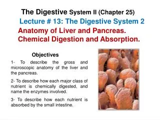
Liver On right under diaphragm, largest organ made up of 4 lobes (left and right, caudate, and quadrate) Hilus (porta hepatis) – underside "entry" point Extremely versatile: Know a few functions? Gall bladder Blood supply to liver Fig 25.20 Fig 25.21 Microscopic anatomy: Liver lobules and triads
Pancreas • Retroperitoneal • Endocrine or exocrine gland? • Common bile duct and pancreatic duct lead to duodenal ampulla and papilla Compare to Figs 25-22 / 23