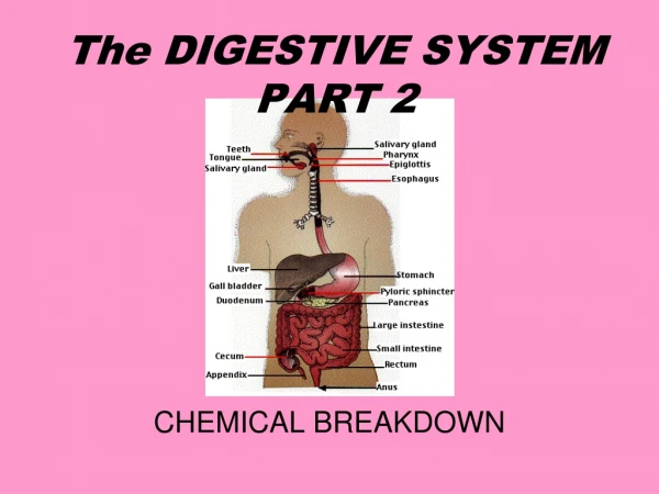
Digestive System Part 2
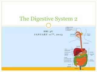
Digestive System Part 2. Anatomy 220. Objectives- Part 1. Describe the relative placement of the following structures & how they fit into the food-digestion pathway : Stomach, liver, gall bladder, pancreas, small intestine

Digestive System Part 2
E N D
Presentation Transcript
Digestive SystemPart 2 Anatomy 220
Objectives- Part 1 • Describe the relative placement of the following structures & how they fit into the food-digestion pathway: • Stomach, liver, gall bladder, pancreas, small intestine • Describe chemical digestion in the stomach listing the secretions, & describing their sources& their basic role in digestion • Describe how the stomach is protected from these secretions
Objectives- Part 2 • Describe the cause of most peptic ulcers • Describe the composition & role of villi • know the organs and ducts associated with the biliary tree & their contributions to the breakdown of fats, and be able to trace the pathway of bile
Objectives- Part 2 · • ··
Functions • Ingestion • Transport • Digestion • Absorption • Compaction • Elimination
Overview • A hollow tube ~ 25 feet (7.5m) long • Changes shape • Several glands secrete into tube
4 Layers • From outer to deep: • Serosa (peritoneum) – CT • Muscular layer (2 parts) • Longitudinal layer • Circular layer • Submucosa • Muscosa
Why doesn’t stomach digest itself? • Mucous from Mucous cells coats and protects
Digestion in the stomach • Two types of digestion: • Chemical caused by the 4 different secretions • Physical caused by contraction of muscular layer. Result is churning and mixing of bolus with gastric secretions
What do you think? • Why does the stomach have an extra layer of muscle (the oblique layer) compared to the rest of the GI tract?
Pyloric Sphincter Location -End of stomach and beginning of duodenum
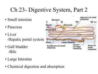
Small Intestine • About 21 ft. long • Divided into three parts: • Duodenum – 1 ft. • Jejunum – 8 ft. • Ileum – 12 ft.
Small Intestine Duodenum Jejunum Ileum
Small Intestines • Most absorption of _________ occur here
Lining of Small Intestines • Inside lining has circular folds called plicae – increase surface area Plicae
Lining of Small Intestines • Entire surface has microscopic _______ called villi (singular villus) Villus
A Villus • A villus has: • epithelium • arteriole • capillary • venuole • lymph duct • Food goes: • across epithelium • into capillary • into venuole • portal v. • liver
What do you think? • Why is the small intestine so much longer than the stomach?
Duodenum • C-shaped • 1 foot long • Mainly on right superior side • Receives secretions from two glands: • Liver – bile (detergent) • Pancreas – bicarbonate and proteolytic enzymes
Pancreatic Duct Common Bile Duct PancreaticDuct
Biliary Tree LIVER GALLBLADDER DUODENUM
Biliary Tree LIVER hepatic ducts GALLBLADDER DUODENUM
Biliary Tree LIVER hepatic ducts common hepatic duct GALL BLADDER DUODENUM
Biliary Tree LIVER hepatic ducts cystic duct common hepatic duct GALLBLADDER DUODENUM
Biliary Tree LIVER hepatic ducts cystic duct common hepatic duct GALLBLADDER common bile duct DUODENUM
Biliary Tree LIVER hepatic ducts cystic duct common hepatic duct GALLBLADDER common bile duct DUODENUM pancreatic duct PANCREAS
Liver Upper Right Quadrant