Chemical Breakdown in Digestive System
280 likes | 327 Vues
Understand the digestion process of carbohydrates, proteins, and lipids in the digestive system. Learn how enzymes break down food in the mouth, stomach, and small intestine. Explore the role of organs like the pancreas, liver, and gallbladder.

Chemical Breakdown in Digestive System
E N D
Presentation Transcript
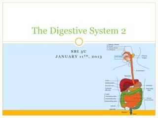
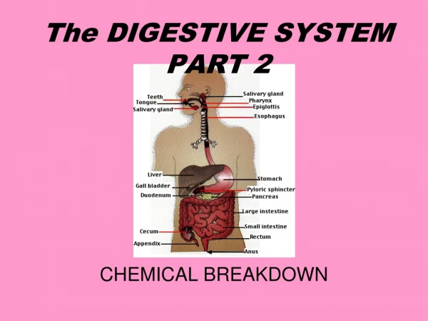
The DIGESTIVE SYSTEMPART 2 CHEMICAL BREAKDOWN
Introduction • Digestion is the chemical breakdown of large food molecules into smaller molecules that can be used by cells. • The breakdown occurs when certain specific enzymes are mixed with the food.
Polysaccharides maltose glucose • Proteins peptides amino acids • Fats fatty acids + glycerol
Carbohydrates, Proteins, Lipids • The process of digestion produces glucose, amino acids, glycerol, and fatty acids. • The energy in glucose is used to produce ATP. (adenosine triphosphate) • The body uses amino acids to construct proteins. • Glycerol and fatty acids can be converted to pyruvate and Acetyl CoA and then enter cellular respiration.
Mouth • Mastication, or chewing, is the first step in the breakdown of complex foodstuffs and serves several functions, including: • breaking large pieces into small pieces, resulting in a massive increase in surface area, which is where digestive enzymes work • softening of food and transformation into a size conducive to swallowing • lubrication of food by impregnating it with saliva
Enzymesin the Mouth • Salivary amylase breaks starch (a polysaccharide) down to maltose (a disaccharide). • Bicarbonate ions in saliva act as buffers, maintaining a pH between 6.5 and 7.5. (pH = measure of acid concentration) • Mucins (mucous) lubricate and help hold chewed food together in a clump called a bolus. • The tongue contains chemical receptors in structures called taste buds. The tongue is muscular and can move food. It pushes food to back where it is swallowed.
Pharynx • The respiratory and digestive passages meet in the pharynx. They separate posterior to the pharynx to form the esophagus (leads to the stomach) and trachea (leads to the lungs). • Swallowing is accomplished by reflexes that close the opening to the trachea. • When swallowing, the epiglottis covers the trachea to prevent food from entering. • In the mouth, food is mixed with saliva and formed into a bolus. • Peristalsis refers to rhythmic contractions that move food in the gut. Peristalsis in the esophagus moves food from the mouth to the stomach.
Stomach • The stomach stores up to 2 liters of food. • Gastric glands within the stomach produce secretions called gastric juice. • The muscular walls of the stomach contract vigorously to mix food with gastric juice, producing a mixture called chyme.
Gastric juice • Pepsinogenis converted to pepsin, which digests proteins. Pepsinogen production is stimulated by the presence of gastrin in the blood (discussed next). • Hydrochloric acid (HCl) converts pepsinogen to pepsin which breaks down proteins to peptides. HCl maintains a pH in the stomach of 2.0. • It also dissolves food and kills microorganisms. • Mucousprotects the stomach from HCl and pepsin.
Secretion of Gastric Juice • Seeing, smelling, tasting, or thinking about food can result in the secretion of gastric juice. • Gastrin is a hormone that stimulates the stomach to secrete gastric juice. • Ulcer • An ulcer is an irritation due to gastric juice penetrating the mucous lining of the stomach or duodenum. It is believed that ulcers are caused by the bacterium Helicobacter pylori, which, can thrive in the acid environment of the stomach. The presence of the bacteria on portions of the stomach lining prevents it from secreting mucous, making it susceptible to the digestive action of pepsin.
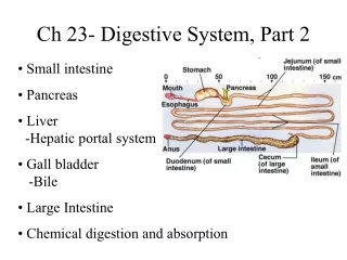
Duodenum • The duodenum is the first part of the small intestine. • Chyme enters through a pyloricsphincter in tiny spurts. • At this point, proteins and carbohydrates are only partially digested and lipid digestion has not begun. • Two ducts enter the duodenum: • one draining the gall bladder and hence the liver, • the other draining the exocrine portion of the pancreas.
Pancreas • The pancreas acts as an exocrine gland by producing pancreatic juice which empties into the small intestine via a duct. • The pancreas also acts as an endocrine gland to produce insulin.
Pancreatic Juice • Pancreatic juice contains sodium bicarbonate which neutralizes the acidic material from the stomach. • Pancreatic amylase digests starch to maltose. • Trypsin andChymotrypsindigest proteins to peptides. Like pepsin (produced in the stomach), they are specific for certain amino acids, not all of them. They therefore produce peptides. • Lipase digests fats to glycerol and fatty acids.
Liver • The liver produces bile which is stored in gallbladder and sent to the duodenum through a duct. • Bile emulsifies fats (separates it into small droplets) using bile acids so they can mix with water and be acted upon by enzymes. • Bile also contains bile pigments. These are the products of the breakdown of hemoglobin removed by the liver from old red blood cells. • The brownish color of the bile pigments imparts the characteristic brown color of the feces.
Other Functions of the Liver • The liver detoxifies blood from intestines that it receives via the hepatic portal vein. • The liver stores glucose as glycogen (animal starch) and breaks down glycogen to release glucose as needed. This storage-release process maintains a constant glucose concentration in the blood (0.1%). If glycogen and glucose run short, proteins can be converted to glucose. • It produces blood proteins. • It destroys old red blood cells and converts hemoglobin from these cells to bilirubin and biliverdin which are components of bile. • Ammonia produced by the digestion of proteins is converted to a less toxic compound (urea) by the liver.
The Importance of the Liver • Glucose is removed and converted into glycogen. • Other monosaccharides are removed and converted into glucose. • Excess amino acids are removed and deaminated. • The amino group is converted into urea. • The residue can then enter the pathways of cellular respiration and be oxidized for energy. • Many nonnutritive molecules, such as ingested drugs, are removed by the liver and, often, detoxified. • The liver serves as a gatekeeper between the intestines and the general circulation. It screens blood reaching it in the hepatic portal system so that its composition when it leaves will be close to normal for the body. • Furthermore, this homeostatic mechanism works both ways. When, for example, the concentration of glucose in the blood drops between meals, the liver releases more to the blood by converting its glycogen stores to glucose (glycogenolysis) • converting certain amino acids into glucose (gluconeogenesis).
Small Intestine • The small intestine is approximately 3 m long. • Like the stomach, it contains numerous ridges and furrows. • In addition, there are numerous projections called villi that function to increase the surface area of the intestine. • Individual villus cells have microvilliwhich greatly increase absorptive surface area. • The total absorptive surface area is equivalent to 500 or 600 square meters. • Each villus contains blood vessels and a lacteal (lymph vessel). • Peptidases and maltase are embedded within the plasma membrane of the microvilli.
Small Intestine - Villi • The crypts at the base of the villi contain • stem cells that continuously divide by • mitosis producing more stem cells • cells that migrate up the surface of the villus while differentiating into • columnar epithelial cells (the majority). They are responsible for digestion and absorption. • goblet cells, which secrete mucus; • endocrine cells, which secrete a variety of hormones;
Peptidases complete the digestion of peptides to amino acids. • Maltasecompletes the digestion of disaccharides. Absorption • Absorption is an important function of the small intestine. • Active transport moves glucose and amino acids into the intestinal cells, then out where they are picked up by capillaries. • Glycerol and fatty acids produced by the digestion of fat enter the villi by diffusion and are reassembled into fat (triglycerides). They combine with proteins and are expelled by exocytosis. They move into the lacteals for transport via the lymphatic system.
Large Intestine • The large intestine is also called the colon. It receives approximately 10 liters of water per day. 1.5 liters is from food and 8.5 liters is from secretions into the gut. 95% of this water is reabsorbed. • The large intestine also absorbs sodium and other ions but it excretes other metallic ions into the wastes. • If water is not absorbed, diarrhea can result, causing dehydration and ion loss. • It absorbs vitamin K produced by colon bacteria. • The last 20 cm of the large intestine is the rectum. Feces is composed of approximately 75% water and 25% solids. One-third of the solids is intestinal bacteria, 2/3’s is undigested materials.
The cecum is a pouch at the junction of the small intestine and large intestine. In herbivorous mammals, it is large and houses bacteria capable of digesting cellulose. In human ancestors, the cecum was larger but has been reduced by evolutionary change to form the appendix. • Polyps are small growths in the epithelial lining of the colon. • They can be benign or cancerous and can be removed individually. • A low-fat, high-fiber diet promotes regularity and is recommended as a protection against colon cancer. • Appendix • The appendix is attached to cecum. • Appendicitis is an infection. The appendix may swell and burst, leading to peritonitis (infection of the abdominal lining).
Ascending colon The ascending colon is on the right side of the abdomen. It is the part of the colon from the cecum to the hepatic flexure (the turn of the colon by the liver). It is retroperitoneal in most humans. In grazing animals the cecum empties into the spiral colon. • Transverse colon The transverse colon is the part of the colon from the hepatic flexure (the turn of the colon by the liver) to the splenic flexure (the turn of the colon by the spleen). The transverse colon hangs off the stomach, attached to it by a wide band of tissue called the mesocolon. The transverse colon is mobile (unlike the parts of the colon immediately before and after it), and is very mobile in the abdomen of some individuals.
Descending colon The descending colon is the part of the colon from the splenic flexure to the beginning of the sigmoid colon. • Sigmoid colon The sigmoid colon is the part of the large intestine after the descending colon and before the rectum. The name sigmoid means S-shaped (see sigmoid). The walls of the sigmoid colon are muscular, and contract to increase the pressure inside the colon, causing the stool to move into the rectum. • Due to the intermittent high pressure within it, the colon can develop pockets calleddiverticuli in its walls. The presence of diverticuli, whether harmful or not, is called diverticulosis. An infection of the diverticuli is called diverticulitis. • Rectum The rectum is the last part of the colon. It holds stool prior to defecation. The last few centimeters of the rectum are lined by tissue which is similar to skin. This area is known as the "social part" of the rectum, since it can distinguish between solid, liquid and gas. That perceptual ability is important in knowing what can be passed appropriately in what circumstance.
Digestion Part 2 FIN