Download
1 / 18
180 likes | 282 Vues
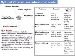
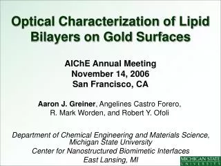
Optical Characterization of Lipid Bilayers on Gold Surfaces. AIChE Annual Meeting November 14, 2006 San Francisco, CA Department of Chemical Engineering and Materials Science, Michigan State University Center for Nanostructured Biomimetic Interfaces East Lansing, MI.

E N D
Optical Characterization of Lipid Bilayers on Gold Surfaces AIChE Annual Meeting November 14, 2006 San Francisco, CA Department of Chemical Engineering and Materials Science, Michigan State University Center for Nanostructured Biomimetic Interfaces East Lansing, MI Aaron J. Greiner, Angelines Castro Forero, R. Mark Worden, and Robert Y. Ofoli
Outline • Motivation and approach • Bilayer lipid membranes » supported bilayer lipid membranes (sBLMs) » tethered bilayer lipid membranes (tBLMs) • Optical characterization » FRAPP • Results • Conclusions
Bilayer Lipid Membranes in Cells • Separates interior of cell from outer environment • A fluid mosaic of phospholipids, proteins, cholesterol, etc. www.anti-age.biz/Glyco-cellular.JPG • Goal: to construct artificial BLMs on substrates to measure protein functionality in a natural setting
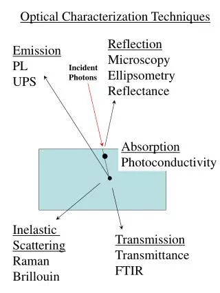
Strategy • Construct biosensor/bioelectronic array devices on gold » measure electrochemical behavior in lipid bilayers • Optically characterize interfaces on semi-transparent gold using FRAPP » measure diffusion coefficient of lipids
Why Gold?? • Advantages » easy to pattern on silicon and glass substrates » easy to make monolayers using thiol-terminated lipids • Disadvantages » quenched fluorescence signal
Supported membranes (sBLMs) Liposome Substrate • Not ideal for ion channel or membrane protein studies » interactions between substrate and protein » lack of sufficient ion reservoir Utility: provides benchmark for evaluating tethered systems
sBLM Lipids 98:2 (mol%) POPC : NBC PC 1-Palmitoyl-2-Oleoyl-sn-Glycero-3-Phosphocholine (POPC) Acyl 06:0 NBD PC
Shutter To Sample Laser Beam Expander Iris Filter Optical Flat #1 Optical Flat #2
FRAPP BLM on gold Objective Laser Light (488 nm) Dichroic Mirror Filter Ronchi Ruling Filter To PMT
FRAPP Photobleach Recovery Monitoring beam : 2 µW(continuous) Photobleaching beam : 500 mW for 400ms D = 200 µm D = 50 µm
Characterization of sBLMs Multi-component diffusion D1 = 5.19 ± 0.58 µm2/s D2 = 0.38 ± 0.15 µm2/s
sBLM w/Cholesterol 68:30:2 (mol%) POPC: Cholesterol: NBC PC Multi-component diffusion D1 = 2.98 ± 0.64 µm2/s D2 = 0.26 ± 0.15 µm2/s
Tethered BLMs Substrate • Appropriate for membrane protein studies » reduces substrate protein interactions » sufficient space for ion reservoir
tBLM Protocol • Monolayer formation on semi-transparent gold (24 hrs) » 1 µmol phosphothioethanol in chloroform • Upper leaflet formed by liposome rupture (1 hr) » 98:2 (mol%) POPC : NBD PC 1,2-Dipalmitoyl-sn-Glycero-3-Phosphothioethanol
Characterization of tBLMs Single-component diffusion D1 = 2.80 ± 0.77 µm2/s
Conclusions • FRAPP can be used to effectively characterize membrane interfaces on biosensor/bioelectronic arrays • Diffusion in tBLMs is comparable to that in sBLMs w/cholesterol • Single component diffusion model fits better for tBLMs on gold » fluorophores preferentially reside in upper leaflet » two component diffusion w/ quenched lower leaflet signal
Acknowledgements • Josh Matter (ChE undergraduate, MSU) • Michigan Technology Tri-Corridor