Refraction - I
540 likes | 1.47k Vues
Learn the basics of refraction, myopia, and its management in ophthalmology. Understand refractive errors, anomalies, and types of myopia like axial and curvature myopia. Gain insight into causes, clinical course, and differences between simple and pathological myopia.

Refraction - I
E N D
Presentation Transcript
Refraction - I Dr Ajai Agrawal Additional Professor Department of Ophthalmology AIIMS Rishikesh
Acknowledgement • Photographs in this presentation are courtesy of Kanski’s Clinical Ophthalmology.
Learning Objectives At the end of the class, students shall be able to • Understand what is refraction. • Have basic knowledge of myopia and its management.
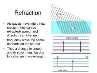
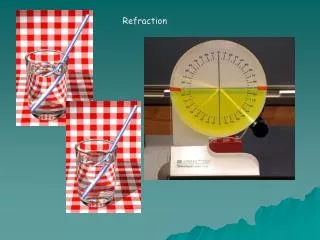
What is Refraction • When rays of light traveling through air enter a denser transparent medium, the speed of the light is reduced and the light rays proceed at a different angle, i.e., they are refracted. • Except when the rays are normal Refraction in Ophthalmology • Methods for evaluating the optical and refractive state of the eye
Emmetropia • Parallel light rays, from an object more than 6 m away, are focused at the plane of the retina when accomodation is at rest. • Clear image of a distant object formed without any internal adjustment of the optics of the eye. • Absence of emmetropia = Ametropia
Progress of refractive state of eye • Birth : +2 to +3 D • 90% of children at age 5 yrsare Hypermetropic • 50% of children at age 16 yrsare Hypermetropic • After the period of growth has passed the refractive state tends to remain stationary, until in old age a further tendency of hypermetropia is evident.
Refractive data in adult • Normal axial length ≈ 24 mm • Change in axial length by 1mm = ±3D • Refraction at corneal surface= +40 to 45(+43)D • Change in Corneal Curvature by 1mm = ±6D • Refraction by unaccomodated lens= +16 to 20(+17)D
Angle kappa (κ) • M = Macula • D= Centre of pupil, on cornea • N = Nodal point M Optic axis D N B κ( FD = Pupillary line FNM = Visual axis F κ = “Between the visual axis and pupillary line, hence roughly corresponds to angle α”.
Anisometropia • Anisometropia is a state in which there is a difference in the refractive errors of the two eyes, i.e., one eye is myopic and the other hyperopic, or both are hyperopic or myopic but to different degrees. • This condition may be congenital or acquired due to asymmetric age changes or disease.
Refractive errors Anomalies of the optical state of the eye Myopia Hypermetropia Astigmatism
What is Myopia ? • Diopteric condition of the eye where parallel incident rays from optical infinity focus anterior to light sensitive layers of retina when accomodation is at rest
Myopia – Optics Emmetropia Diverging lens
Optics of Myopic eye • Far point is at a finite distance inversely proportional to the degree of myopia • Weakest concave lens that diverges rays just sufficiently to focus them at the retina is to be used • Poor visual acuity is compensated to some extent by enlarged image size due to the nodal point being further from the retina
Causes of Myopia • The causes of myopia are not known. • Epidemiological correlation suggest... • lengthy periods of close work are probably a contributory factor • there is some genetic predisposition to myopia and its severity
Types of myopia Axial Curvature Index Positional
Axial Myopia • AP diameter increased to 25.5 to 32.5 mm • 90-95% cases • There may be… • pseudoproptosis resulting from the abnormally large anterior segment, • a peripapillary myopic crescent from an exaggerated scleral ring, • posterior staphyloma
Curvature Myopia • Corneal curvature steeper than average, e.g., keratoconus, • Radius <7-8.5 mm (normal); 1 mm=6 D • Lens curvature is increased • moderate to severe hyperglycemia (intumescence) lenticonus (ant/post) spasm of accomodation spherophakia
Index Myopia • Increased index of refraction in early to moderate nuclear sclerotic cataracts in the elderly. • Many people find themselves ultimately able to read without glasses or having gained “second sight.” • Decrease in refractive index of cortex – diabetic myopia
Positional Myopia • Anterior movement of the lens is often seen after glaucoma surgery and will increase the myopic error in the eye. • Axial myopia of buphthalmos is countered to a large extent due to posterior displacement of lens-iris diaphragm and flattening of the cornea
Clinical course Simple Pathological
Simple Myopia • Rarely present at birth, but often begins to develop as the child grows. • Usually detected by age 9 or 10 years in the school vision tests • May increase during the years of growth, stabilizing around the mid-teens, usually at about 5 D or less.
Pathological Myopia • 2-3% population • Increases by as much as 4 D/yr • Usually stabilizes at about age 20 years and frequently results in myopia – 10 to 20 D. • If progress is rapid from age 15-20, likely to reach 20-30 dioptres • Commoner in women, Jews and Japanese
Pathological Myopia-Etiology • Developmental defect affecting posterior segment • Retina grows extensively stretching sclera • Adjuvants- growth influences during puberty and physical debility • Excessive convergence- stretching
Pathological Myopia • Associated vitreous floaters, liquefaction, posterior staphyloma and chorioretinal changes. • Degeneration is not necessarily comparable with degree of myopia • Genetic predisposition in offspring as per laws of recessive Mendelian inheritance – if both parents affected, close supervision needed
School/ Physiologic/Pseudo-Myopia • ≤ 2D • Excessive near work causing accomodative spasm • Inherited predisposition-more in Orientals and Jews
Symptoms • Blurred distance vision. • Squinting to sharpen distance vision by attempting a pinhole effect through narrowing of the palpebral fissures. • Eye strain seen in patients with uncorrected low myopic errors • Closer working distance at near that typically gets closer and closer as the person sustains working at near. • Delayed dark adaptation • Floaters, photopsiae • Visual deterioration
Signs • Small eyeball • Smaller cornea • Shallow anterior chamber predisposes to angle closure glaucoma since size of lens is normal • Apparent divergent squint
Clinical Signs – Apparent convergent squint • The problem begins at near and spreads to distance leading to a cascade of changes in the findings over time • Results usually in apparent convergent squintdue to excess convergence
Clinical Signs – True divergent squint • Excess convergence for near work disorients accommodation which may increase causing ciliary spasm or • more frequently, attempt at convergence is given up, its latent insufficiency causing muscular imbalance till • advantage of binocular vision is given up, one eye is relied upon for vision while the other deviates outwards causing true divergent squint
Pathology • Eye appears large and prominent – pseudoproptosis • Deep anterior chamber • Large, sluggish pupil • Post segment sclera is thinned up to 25% of normal • Post vitreous detachment – Weiss ring • Liquefaction – muscae volitantes, large floaters
Fundus • Atrophy of retina and choroid – depigmentation • Tigroid fundus with prominent choroidal vessels • Patches of choroidal atrophy surrounded by pigment associated with haemorrhages • Atrophic patch at macula associated with loss of central vision
Fundus • Appearance of dark pigmented area at macula-Foster- Fuch’s fleck – rare, sudden, proliferation of pigmentary epithelium with intra-choroidal haemorrhage or thrombosis • Macular bunches of dilated capillaries or aneurysms • Myopic crescent – temporal or annular • Nasal supertraction crescent
Posterior staphyloma • Herniation of posterior pole • Crescentric shadow 2-3 DD temporal to disc, • Sudden kinking of retinal vessels as they dip over the edges, • Gross atrophy
Peripheral Degenerations Not requiring prophylaxis: Paving stone
Predisposing Degenerations Lattice, snailtrack, retinoschisis, white without pressure Retinoschisis Snailtrack
Lattice degeneration Figure:
Complications • Atrophy – scotomata – macular most incapacitating • Vitreous degeneration + floaters • Tears + haemorrhages • Detachment – post traumatic or spontaneous associated with peripheral degenerations due to vitreous adhesion • Lenticular opacities, esp. posterior cortical • Open angle glaucoma Horseshoe Tear
Night myopia • Manifest in reduced illumination • ~ 0.5 D • Cone-rod shift in retina, pupillary dilatation, ciliary muscle activity • If night vision appears seriously impaired, appropriate correction may be given
Treatment • Optical correction after subjective and objective refraction • Spectacles • Contact lens (including Orthokeratology) • Visual hygiene • Refractive surgery • LASIK o LASEK • Wavefront Lasik o Clear lens Extraction • Phakic IOL o ICRS • Pharmacological intervention
Myopia – Optics Diverging lens
Cycloplegic Refraction • Cycloplegia is the employment of pharmaceutical agents to paralyze the ciliary muscle temporarily to stabilize the accommodative reflex of the eye so that a definitive end point may be measured. • Benefit of relaxing the accommodative tone is especially important in young individuals. • Cycloplegic + Mydriatic = Relaxes accomodation + dilates pupil for better reflex
Visual Hygiene • Proper illumination • Proper posture • Clear print • Better contrast • Avoid ocular fatigue • Proper occupation in case of degenerative myopia • May need special institutions if low vision dictates
Summary • Refraction is a method for evaluating the optical and refractive state of the eye. • Myopia is a diopteric condition of the eye where parallel incident rays from optical infinity focus anterior to light sensitive layers of retina when accomodation is at rest. • Myopia is corrected by concave lenses prescribed after cycloplegic refraction.