Download
1 / 72
730 likes | 955 Vues
GENETIC DISORDERS. Introduction of the Human Genome. The complete set of instructions تعليمات توصيات for making an organism is called its genome . It contains the master blueprint مخطط أو برنامج عمل for all cellular structures and activities for the lifetime of the cell or organism.

E N D
Introduction of the Human Genome • The complete set of instructions تعليمات توصيات for making an organism is called its genome. • It contains the master blueprint مخطط أو برنامج عملfor all cellular structures and activities for the lifetime of the cell or organism. • Found in every nucleus of a persons many trillions of cells, the human genome consists of tightly coiled threads of deoxyribonucleic acid (DNA) and associated protein molecules, organized into structures called chromosomes. • For each organism, the components of these slender threads encode يحول الى رموز تلغرافية all the information necessary for building and maintaining life, from simple bacteria to remarkably complex human beings. • Understanding how DNA performs this function requires some knowledge of its structure and organization.
DNA • In humans, as in other higher organisms, a DNA molecule consists of two strands that wrap around each other to resemble a twisted ladder whose sides, made of sugar and phosphate molecules, are connected by rungsدرجة سلم of nitrogen- containing chemicals called bases. • Each strand is a linear arrangement of repeating similar units called nucleotides, which are each composed of one sugar, one phosphate, and a nitrogenous base. Four different bases are present in DNA: adenine (A), thymine (T), cytosine (C), and guanine (G). • The particular order of the bases arranged along the sugar- phosphate backbone is called the DNA sequence; the sequence specifies the exact genetic instructions required to create a particular organism with its own unique traits. • The two DNA strands are held together by weak bonds between the bases on each strand, forming base pairs (bp). • Genome size is usually stated as the total number of base pairs; the human genome contains roughly 3 billion bp.
Each time a cell divides into two daughter cells, its full genome is duplicated; for humans and other complex organisms, this duplication occurs in the nucleus. • During cell division the DNA molecule unwindsيفك and the weak bonds between the base pairs break, allowing the strands to separate. • Each strand directs the synthesis of a complementary new strand, with free nucleotides matching up with their complementary bases on each of the separated strands. • Strict base- pairing rules are adhered to adenine will pair only with thymine (an A- T pair) and cytosine with guanine (a C- G pair). • Each daughter cell receives one old and one new DNA strand. • The cells adherence to these base- pairing rules ensures that the new strand is an exact copy of the old one, which minimizes the incidence of errors (mutations).
Chromosome Region of euchromatin with activated genes Nucleosome DNA
Deoxyribonucleic acid (DNA). Base pairings between a purine and pyrimidine base link thetwo polynucleotide strands of the DNA double helix.
Genes • Each DNA molecule contains many genes = the basic physical and functional units of heredity. • A gene is a specific sequence of nucleotide bases, whose sequences carry the information required for constructing proteins, which provide the structural components of cells and tissues as well as enzymes for essential biochemical reactions. • The human genome is estimated to comprise more than 25,000 genes. • Human genes vary widely in length, often extending over thousands of bases, but only about 10% of the genome is known to include the protein- coding sequences (exons) of genes. • Interspersed within many genes are intron sequences, which have no coding function. • The balance of the genome is thought to consist of other noncoding regions (such as control sequences and intergenic regions), whose functions are obscure. • All living organisms are composed largely of proteins; humans can synthesize at least 100,000 different kinds. • Proteins are large, complex molecules made up of long chains of subunits called amino acids. • Twenty different kinds of amino acids are usually found in proteins. • Within the gene, each specific sequence of three DNA bases (codons) directs the cells protein- synthesizing machinery to add specific amino acids
For example, the base sequence ATG codes for the amino acid methionine. • Since 3 bases code for 1 amino acid, the protein coded by an average- 3 sized gene (3000 bp) will contain 1000 amino acids. • The genetic code is thus a series of codons that specify which amino acids are required to make up specific proteins. • The protein- coding instructions from the genes are transmitted indirectly through messenger ribonucleic acid (mRNA), a transient intermediary molecule similar to a single strand of DNA. • For the information within a gene to be expressed, a complementary RNA strand is produced (a process called transcription) from the DNA template in the nucleus. • This mRNA is moved from the nucleus to the cellular cytoplasm, where it serves as the template for protein synthesis. • The cells protein- synthesizing machinery then translates the codons into a string of amino acids that will constitute the protein molecule for which it codes. • In the laboratory, the mRNA molecule can be isolated and used as a template to synthesize a complementary DNA (cDNA) strand, which can then be used to locate the corresponding genes on a chromosome map.
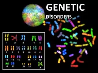
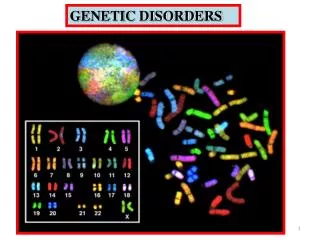
Chromosomes • The 3 billion bp in the human genome are organized into 24 distinct physically separate chromosomes. • All genes are arranged linearly along the chromosomes. • The nucleus of most human cells contains 2 sets of chromosomes, 1 set given by each parent. • Each set has 23 single chromosomes; 22 autosomes and a sex chromosome (X or Y). • (A normal female has a pair of X chromosomes; while a male has an X and Y pair.) • Chromosomes contain roughly equal parts of protein and DNA; chromosomal DNA contains an average of 150 million bases. • DNA molecules are among the largest molecules now known. • Chromosomes can be seen under a light microscope and, when stained with certain dyes, reveal a pattern of light and dark bands reflecting regional variations in the amounts of A and T vs G and C. • Differences in size and banding pattern allow the 24 chromosomes to be 4 distinguished from each other, an analysis called a karyotype.
The structure of a chromosome after DNA replication. At this stage, a chromosome consists of two identical strands, or chromatids. Centromere chromatide DNA Histone
A few types of major chromosomal abnormalities, including missing or extra copies of a chromosome or gross breaks and rejoinings (translocations), can be detected by microscopic examination; Downs syndrome, in which an individual's cells contain a third copy of chromosome 21, is diagnosed by karyotype analysis. • Most changes in DNA, however, are too subtle to be detected by this technique and require molecular analysis. • These subtle DNA abnormalities (mutations) are responsible for many inherited diseases such as cystic fibrosis and sickle cell anemia or may predispose an individual to cancer, major psychiatric illnesses, and other complex diseases.
DISEASES • GENETIC • ENVIRONMENTAL • BOTH
MUTATIONS • PERMANENT change in DNA 1- GENOME MUTATION: (whole chromosome) 2- CHROMOSOME MUTATION: (visible chromosome change) 3- GENE MUTATION: (may, and often, result in a single base error)
GENE MUTATION 1- DELETION OF A SINGLE BASE 2- SUBSTITUTION OF A SINGLE BASE
GENE MUTATION 1- POINT MUTATION within a coding sequence: VAL-GLU 2- MUTATIONS in NON-coding sequences defective transcription 3- DELETIONS/INSERTIONS frame shift mutation, involvement is NOT a multiple of 3 4- Tri-nucleotide REPEATS, e.g., CGG repeats many times in fragile X syndrome
GENE MUTATIONS 1- INTERFERE with protein synthesis 2- SUPPRESS transcription, DNARNA 3- PRODUCE abnormal mRNA 4- DEFECTS carried over into TRANSLATION 5- ABNORMAL proteins WITHOUT impairing syntheses
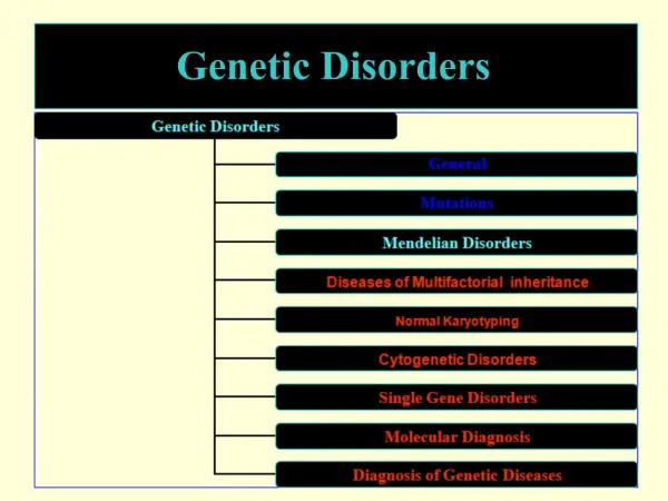
GENETIC DISORDERS 1- SINGLE gene mutations, following classical MENDELIAN inheritance patterns 2- MULTIFACTORIAL inheritance 3- CHROMOSOMAL disorders
MENDELIAN inheritance patterns 1- AUTOSOMAL DOMINANT 2- AUTOSOMAL RECESSIVE 3- SEX-LINKED (recessive), involving “X” chromosome
AUTOSOMAL DOMINANT • Disease is in HETEROZYGOTES • NEITHER parent may have the disease (NEW mut.) • REDUCED PENETRANCE (env?, other genes?) • VARIABLE EXPRESSIVITY (env?, other genes?) • May have a DELAYED ONSET • Usually result in a REDUCED PRODUCTION or INACTIVE protein
AUTOSOMAL DOMINANT • HUNTINGTON DISEASE • NEUROFIBROMATOSIS • MYOTONIC DYSTROPHY • TUBEROUS SCLEROSIS • POLYCYSTIC KIDNEY • HEREDITARY SPHEROCYTOSIS • VON WILLEBRAND DISEASE • MARFAN SYNDROME • EHLERS-DANLOS SYNDROMES (some) • OSTEOGENESIS IMPERFECTA • ACHONDROPLASIA • FAMILIAL HYPERCHOLESTEROLEMIA • ACUTE INTERMITTENT PORPHYRIA
AUTOSOMAL DOMINANT PEDIGREE النسب 1) BOTH SEXES INVOLVED 2) GENERATIONS NOT SKIPPED aa Aa
AUTOSOMAL RECESSIVE • Disease is in HOMOZYGOTES • More UNIFORM expression than AD • Often COMPLETE PENETRANCE • Onset usually EARLY in life • NEW mutations rarely detected clinically • Proteins show LOSS of FUNCTION • Include ALL inborn errors of metabolism • MUCH more common that autosomal dominant
AUTOSOMAL RECESSIVE • CF • PKU • GALACTOSEMIA • HOMOCYSTINURIA • LYSOSOMAL STORAGE • Α-1 ANTITRYPSIN • WILSON DISEASE • HEMOCHROMATOSIS • GLYCOGEN STORAGE DISEASES Hgb S THALASSEMIAS CONG. ADRENAL HYPERPLASIA EHLERS-DANLOS (some) ALKAPTONURIA NEUROGENIC MUSC. ATROPHIES FRIEDREICH ATAXIA SPINAL MUSCULAR ATROPHY
AUTOSOMAL RECESSIVE PEDIGREE 1) BOTH SEXES INVOLVED 2) GENERATIONS SKIPPED
SEX (“X”) LINKED • MALES ONLY • HIS SONS are OK • ALL his DAUGHTERS are CARRIERS • The “Y” chromosome is NOT homologous to the “X”, i.e., the concept of dominant/recessive has no meaning here • HETEROZYGOUS FEMALES have no phenotypic expression (carriers)
SEX (“X”) LINKED • DUCHENNE MUSCULAR DYSTROPHY • HEMOPHILIA , A and B • G6PD DEFICIENCY • AGAMMAGLOBULINEMIA • WISKOTT-ALDRICH SYNDROME • DIABETES INSIPIDUS • LESCH-NYHAN SYNDROME • FRAGILE-X SYNDROME
SEX LINKED PEDIGREE 1) MALES ONLY 2) GENERATION SKIPPING DOESN’T MATTER
SINGLE GENE DISORDERS • 1- ENZYME DEFECT (Most of them, e.g., PKU) • Accumulation of substrate • Lack of product • Failure to inactivate a protein which causes damage • 2- RECEPTOR/TRANSPORT PROTEIN DEFECT (Familial Hypercholesterolemia) • 3- STRUCTURAL PROTEIN DEFECT (Marfan, Ehl-Dan) • Structure • Function • Quantity • 4- ENZYME DEFECT WHICH INCREASES DRUG SUSCEPTIBILITY: G6PDPrimaquine
STRUCTURAL PROTEIN DEFECTS • 1- Marfan Syndrome • Fibrillin-1 defect • Tall, dislocated lens, aortic arch aneurysms, etc. • Abraham Lincoln?, Osama bin-Laden • 2- Ehlers- Danlos Syndromes (AD, AR) • Multiple (6?) different types • Classical, Hypermob., Vasc., KyphoSc., ArthChal., Derm • Various collagen defects • Hyperelastic skin, hyperextensible joints
RECEPTOR PROTEIN DEFECTS 1- FAMILIAL HYPERCHOLESTEROLEMIA • LDL RECEPTOR defect • Cholesterol TRANSPORT across liver cell impaired • ergo, CHOLESTEROL BUILDUP IN BLOOD 2- “Scavenger System” for CHOL kicks in, i.e., MACROPHAGES • YOU KNOW THE REST OF THE STORY • YOU KNOW WHY MACROPHAGES are “FOAMY”
ENZYME DEFICIENCIES • BY FAR, THE LARGEST KNOWN CATEGORY • SUBSTRATE BUILDUP • PRODUCT LACK • SUBSTRATE could be HARMFUL • LYSOSOMAL STORAGE DISEASES comprise MOST of them
LYSOSOMAL STORAGE DISEASES • GLYCOGEN STORAGE DISEASES • SPHINGOLIPIDOSES (Gangliosides) • SULFATIDOSES • MUCOPOLYSACCHARIDOSES • MUCOLIPIDOSES • OTHER • Fucosidosis, Mannosidosis, Aspartylglycosaminuria • WOLMAN, Acid phosphate deficiency
GLYCOGEN STORAGE DISEASES • MANY TYPES (at least 10) • Type 2 (Pompe), von Gierke, McArdle, most studied and discussed, and referred to • Storage sites: Liver, Muscle, Heart
SPHINGOLIPIDOSES • MANY types, Tay -Sachs most often referred to • GANGLIOSIDES are ACCUMULATED • Ashkenazi Jews (1/30 are carriers) • CNS neurons a site of accumulation • CHERRY RED spot in Macula
SULFATIDOSES • MANY types, but the metachromatic leukodystrophies (CNS), Krabbe, Fabry, Gaucher, and Niemann -Pick (A and B) are most commonly referred to • SULFATIDES, CEREBROSIDES, SPHINGOMYELIN are the accumulations
NIEMANN-PICK • TYPES A, B, C • SPHINGOMYELIN • MASSIVE SPLENOMEGALY • ALSO in ASHKANAZI JEWS • OFTEN FATAL in EARLY LIFE, CNS, ORGANOMEGALY
GAUCHER DISEASE • GLUCOCEREBROSIDE BUILDUP • 99% are type I, NO CNS involvement • ALL MACROPHAGES, liv, spl, nodes, marrow
MUCOPOLYSACCHARIDOSES • HURLER/HUNTER, for I and II, respectively • DERMATAN sulfate, HEPARAN sulfate buildup • coarse facial features • clouding of the cornea • joint stiffness • mental retardation • URINARY EXCRETION of SULFATES COMMON
OTHER LYSOSOMAL STORAGE DIS. • FUCOSIDOSIS • MANNOSIDOSIS • ASPARTYLGLYCOSAMINURIA • WOLMAN (CHOL., TRIGLYCERIDES) • ACID PHOSPHATE DEFICIENCY (PHOS. ESTERS)
ALCAPTONURIA • NOT a LYSOSOMAL ENZYME DISEASE • FIRST ONE TO BE DESCRIBED • HOMOGENTISIC ACID • HOMOGENTISIC ACID OXIDASE • BLACK URINE • BLACK NAILS (OCHRONOSIS), SKIN • BLACK JOINT CARTILAGE (SEVERE ARTHRITIS)
NEUROFIBROMATOSIS • 1 and 2 • 1-von Recklinghausen • 2- “acoustic” neurofibromatosis • 1 • Neurofibromas, café-au- lait, Lisch nodules
NEUROFIBROMATOSIS • 1 and 2 • 1-von Recklinghausen • 2- “acoustic” neurofibromatosis • 2 • Bilateral acoustic neuromas and multiple meningiomas
MULTIFACTORIAL INHERITANCE • Multi-”FACTORIAL”, not just multi-GENIC • “SOIL” theory • Common phenotypic expressions governed by “multifactorial” inheritance • Hair color • Eye color • Skin color • Height • Intelligence • Diabetes, type II
FEATURES of multifactorial inheritance • Expression determined by NUMBER of genes • Overall 5% chance of 1st degree relatives having it • Identical twins >>>5%, but WAY less than 100% • This 5% is increased if more children have it • Expression of CONTINUOUS traits (e.g., height) vs. DISCONTINUOUS traits (e.g., diabetes)
“MULTIFACTORIAL” DISORDERS • Cleft lip, palate • Congenital heart disease • Coronary heart disease • Hypertension • Gout • Diabetes • Pyloric stenosis • MANY, MANY, MANY, MANY MORE
KARYOTYPING • Defined as the study of CHROMOSOMES • 46 = (22x2) + X = Y • Conventional notation is “46,XY” or “46,XX” • G( iemsa )-banding, 500 bands per haploid recognizable • Short (“p”- etit) arm = p, other (long) arm = q