Prostate Pathology
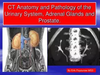
Prostate Pathology. Shaesta Naseem 3-4-2013. Prostate gland. Prostate weighs 20 grams in normal adult Retroperitoneal organ , encircling the neck of bladder and urethra Divided into distinct zones or regions: the peripheral, central, and transitional zones


Prostate Pathology
E N D
Presentation Transcript
Prostate Pathology ShaestaNaseem 3-4-2013
Prostate gland • Prostate weighs 20 grams in normal adult • Retroperitoneal organ ,encircling the neck of bladder and urethra • Divided into distinct zones or regions: the peripheral, central, and transitional zones • Histologically the prostate is composed of glands lined by two layers of cells: a basal layer of low cuboidal epithelium covered by a layer of columnar secretory cells
Zones of the Prostate • Peripheral • Central • Transitional • Most hyperplasias arise in the transitional zone, • Most carcinomas originate in the peripheral zone.
The normal histologic appearance of prostate glands and surrounding fibromuscularstroma A small pink concretion (typical of the corpora amylaceaseen in benign prostatic glands) appears in the gland just to the left of center. Note the well-differentiated glands with tall columnar epithelial lining cells. These cells do not have prominent nucleoli.
Benign Prostatic Hyperplasia(BPH) • Extremely common lesion in men over age 50 (20% in men over age 40,up to 70% by age 60 ,and 90% by age 70) • Hyperplasia of glands and stroma • Fairly large ,well defined nodules • Related to the action of androgen • DHT , Dihydrotestesterone is the ultimate mediator for prostatic growth • Prepubertal castration prevents BPH
BPH , Morphology • The prostate weighs between 60 and 100 grams( normal 20 gm) • Almost exclusively in the transitional zoneof the prostate gland • Nodules ,vary in color and consistency
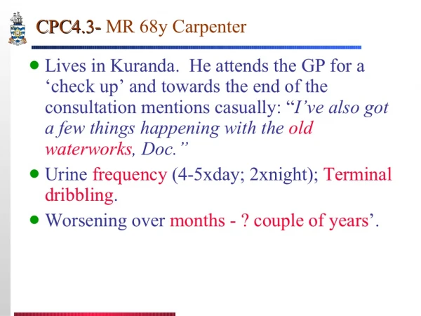
BPH Clinical features • The increased size of the gland, and the smooth muscle-mediated contraction of the prostate cause uretheral obstruction. • The increased resistance to urinary outflow leads to bladder hypertrophy or diverticulum and distension. • The inability to empty the bladder completely creates a reservoir of residual urine common source of infection. • Patients experience increased urinary frequency, nocturia, difficulty in starting and stopping the stream of urine, overflow dribbling, dysuria (painful micturition), and have an increased risk of developing bacterial infections of the bladder and kidney.
BPH, Morphology • The hallmark of BPH is nodularity due to glandular proliferation or dilation and to fibrous or muscular proliferation • Aggregation of small to large to cystically dilated glands • Needle biopsy doesn’t sample the transitional zone where BPH occur
A normal prostate gland is about 3 to 4 cm in diameter. This prostate is enlarged due to prostatic hyperplasia, which appears nodular. Thus, this condition is termed either BPH (benign prostatic hyperplasia) or nodular prostatic hyperplasia
Nodules appear mainly in the lateral lobes. Such an enlarged prostate can obstruct urinary outflow from the bladder causing dysuria and hematuria and lead to an obstructive uropathy or bladder diverticulum
The enlarged prostate gland seen here not only has enlarged lateral lobes, but also a greatly enlarged median lobe that obstructs the prostatic urethra. • This led to obstruction with bladder hypertrophy, as evidenced by the prominent trabeculation of the bladder wall seen here from the mucosal surface. • Obstruction with stasis also led to the formation of the yellow-brown calculus (stone).
BPH can involve both glands and stroma, though the former is usually more prominent. Here, a large hyperplastic nodule of glands is seen. The glands are well-differentiated and still have some intervening stroma. The small laminated pink concretions within the glandular lumens are known as corpora amylacea.
Carcinoma Dysplasia Lumen Severe Microinvasive Mild Moderate In situ Secretory cells Normal Basal Cells Low Grade High Grade Basal Membrane Prostatic Intra-epithelial Neoplasia Prostatic Intraepithelial Neoplasia (PIN) • PIN consists of architecturally benign prostatic acini lined by cytologically atypical cells with prominent nucleoli • PIN is an intermediate lesion between normal and invasive cancer and is considered as predisposing factor and precursor of prostatic adenocarcinoma.
Prostatic Adenocarcinoma • Adenocarcinoma of the prostate is the most common form of cancer in men • Second leading cause of cancer death • Disease of men over age 50 • More prevalent among blacks in the USA Etiology Several risk factors : • Age , race, family history ,hormone level ,and environmental influences . • Androgen are believed to play a role in the pathogenesis
Prostatic Adenocarcinoma , Morphology • 70% arises in the peripheral zone of the gland • Palpablein rectal exam • cross-section of the prostate the neoplastic tissue is gritty and firm
Prostatic adenocarcinoma. • Irregular yellowish nodules. • Prostate glands containing adenocarcinoma are not necessarily enlarged. • Adenocarcinoma may also coexist with hyperplasia. • However, prostatic hyperplasia is not a premalignant lesion
At high magnification, the neoplastic glands of prostatic adenocarcinoma are still recognizable as glands, but there is no intervening stromaand the nuclei are hyperchromatic.
Poorly differentiated prostatic adenocarcinoma demonstrates cells with nucleoli and mitotic figures.
Prostatic Adenocarcinoma,Microscopic • The glands are typically smaller than benign glands and are lined by a single uniform layer of cuboidalor low columnar epithelium. • In contrast to benign glands, prostate cancer glands are more crowded, and characteristically lack branching and papillary infolding. • The outer basal cell layer typical of benign glands is absent. • Nuclei are large and often contain one or more large nucleoli • Peri-neural invasion is common and typical
Spread of prostatic Adenocarcinoma • By direct local invasion and through blood stream and lymph • Local extension most commonly involves the seminal vesicles and the base of the urinary bladder • Hematogenousextension occurs chiefly to the bones. • The bony metastasis are typically osteoblastic
Prostatic Adenocarcinoma Grading and Staging • Gleason grading system is the best known for grading • Five grades on the basis of glandular pattern and degree of differentiation as seen under low magnification • Grading is of particular important in prostate cancer ,because it is the best marker ,along with the stage ,for predicting prognosis • Staging in prostate cancer depends on the TNM system
Prostatic Adenocarcinoma,Clinical Course • Microscopic cancers are asymptomatic, discovered incidentally • Patients with clinically localized disease do not have urinary symptoms • Most arise peripherally ,away from urethra ,therefore ,urinary symptoms occur late
Prostatic Adenocarcinoma ,Clinical Course • Careful digital exam may detect some early cancers • PSA (Prostate Specific Antigen) has been used in the diagnosis and management of prostate cancer • PSA is organ specific but not cancer specific • Could be increased in BPH
Prostatic Adenocarcinoma, Treatment • Surgery ,radiotherapy ,and hormonal therapy • 90% of treated patients expected to live for 15 years • Currently the most acceptable treatment for clinically localized cancer is radical surgery • Locally advanced cancers can be treated by radiotherapy • Hormonal therapy (Anti-androgen therapy) could induce remission .