Download
1 / 19
190 likes | 389 Vues
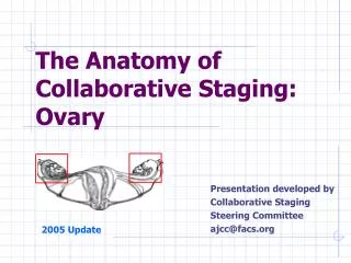
The Anatomy of Collaborative Staging: Ovary. Presentation developed by Collaborative Staging Steering Committee ajcc@facs.org. 2005 Update. Structures of Female Pelvis. Ovarian Cancer. Collaborative Stage fields Tumor Size--standard Extension TS/Ext Eval--standard Lymph Nodes

E N D
The Anatomy of Collaborative Staging: Ovary Presentation developed by Collaborative Staging Steering Committee ajcc@facs.org 2005 Update
Ovarian Cancer • Collaborative Stage fields • Tumor Size--standard • Extension • TS/Ext Eval--standard • Lymph Nodes • LN Eval--standard • LN Pos--standard • LN Exam--standard • Mets at Dx • Mets Eval--standard • Site-specific Factor 1--CA-125 • Site-specific Factors 2-6--not applicable
CS Extension--Notes • Ascites • Positive ascites changes stages I and II to IC and IIC. • Disregard negative ascites or ascites, NOS • Pelvic organs* coded to 50-65 (FIGO Stage II) • Adnexa, bladder (including serosa), uterine ligaments, cul de sac, fallopian tubes, parametrium, pelvic peritoneum, pelvic wall, rectum, sigmoid colon, ureter, uterus, uterine serosa • Abdominal organs* coded to 70-75 (FIGO III) • Abdominal mesentery, diaphragm, gallbladder, infracolic omentum, kidneys, large intestine except rectum and sigmoid, peritoneal surface of liver, omentum, pancreas, pericolic gutter, peritoneum, NOS, small intestine, spleen, stomach, ureters *Involvement may be direct or discontinuous
CS Extension--Notes • Liver parenchymal metastases are coded in Mets at Dx. Implants (discontinuous metastases) Other names: seeding, salting, studding, talcum powder appearance • Determine whether implants are in pelvis (code 60-64) or abdomen (code 70-73) or unspecified (code 75) • Implants outside the pelvis must be microscopically confirmed and coded by size 70 Microscopic only 71 Macroscopic < 2 cm 72 Macroscopic > 2 cm 73 Mets outside pelvis, size not stated 75 Peritoneal implants, NOS [not stated as pelvic or abdominal]
CS Extension Codes (FIGO I) 10 One ovary, capsule intact, no tumor on surface, negative ascites (FIGO IA) 20 Both ovaries, capsules intact, no tumor on surface, negative ascites (FIGO IB) Source: TNM Atlas, 3rd ed. 2nd rev., by B. Spiessl et al. Springer Verlag 1992.
CS Extension Codes (FIGO IC) One or both ovaries 35 Capsule ruptured 36 Tumor on surface 41 One or both ovaries positive ascites or washings Source: TNM Atlas, 3rd ed. 2nd rev., by B. Spiessl et al. Springer Verlag 1992.
CS Extension Codes 50-52 (FIGO IIA) Extension to or implants on: (negative ascites/washings) 50 Adnexa/tubes, ipsilateral 52 Adnexa/tubes, contralateral 52 Uterus Source: TNM Atlas, 3rd ed. 2nd rev., by B. Spiessl et al. Springer Verlag 1992.
CS Extension Codes 60, 61 (FIGO IIB) Extension to or implants on: (negative ascites) 60 Other pelvic structures* (ipsilateral) 61 Other pelvic structures* (contralateral) * ligaments, mesovarium, pelvic wall, adjacent peritoneum Source: TNM Atlas, 3rd ed. 2nd rev., by B. Spiessl et al. Springer Verlag 1992.
CS Extension Codes 62-64 (FIGO IIC) Tumor confined to pelvis with positive ascites 62 Ipsilateral (50 and/or 60) 63 Contralateral (52 and/or 61) 64 Other pelvic structures Source: TNM Atlas, 3rd ed. 2nd rev., by B. Spiessl et al. Springer Verlag 1992.
CS Extension Code 70-75 (FIGO III) 70 Microscopic peritoneal implants (FIGO IIIA) 72 Macroscopic peritoneal implants > 2 cm (FIGO IIIC) 71 Macroscopic peritoneal implants < 2 cm (FIGO IIIB) Source: TNM Atlas, 3rd ed. 2nd rev., by B. Spiessl et al. Springer Verlag 1992.
Liver Involvement CS Extension codes 70-73: Tumor on capsule or surface of liver (FIGO III) Mets at Dx code 40: Metastasis inside liver (parenchymal) (FIGO IV) Source: TNM Atlas, 3rd ed. 2nd rev., by B. Spiessl et al. Springer Verlag 1992.
CS Lymph Nodes--Notes Code distant nodes in Mets at Dx. If there is a statement that “adnexa palpated” with no mention of lymph nodes, assume lymph nodes are not involved (code 00). If exploratory or definitive surgery with no mention of nodes, assume nodes are negative (00). Regional nodes include bilateral and contralateral involvement of named nodes.
CS Lymph Nodes Code 10 1 Hypogastric (internal iliac) Obturator 2 Common iliac 3 External iliac Code 12 4 Lateral sacral Code 20 5 Para-aortic Code 30 6 Inguinal 40 10 + 20 42 [12 or 30] + [10 or 20] Source: TNM-Interactive, UICC, 1998
CS Mets at Dx 10 Distant nodes, NOS 40 Distant metastases except nodes Liver parenchyma Pleural effusion with positive cytology FIGO Stage IVB 50 Distant mets plus distant nodes 99 Unknown, not assessed, not documented
Site-Specific Factor 1 CA-125 • Tumor marker for ovarian cancer; monitors for disease progression • Also called cancer antigen 125 or carbohydrate antigen 125 • Normal range < 35 U/ml (SI: < 35 kU/L) • Code the lowest pre-treatment test • 000 Test not done • 010 Positive/elevated • 020 Negative/normal; within normal limits • 030 Borderline; undetermined whether positive • or negative • 080 Ordered but results not in chart • 999 Unknown, no information, not documented
Ovarian CancerCase Study 1: Limited to ovaries • Enlarged right ovary on PE; mass confirmed by ultrasound. No further tests. Laparotomy, RSO, and random biopsies: tumor confined to ovary, capsule intact. All bx and washings negative • Tumor size 999 Size not stated • Extension 10 One ovary involved, capsule intact • TS/Ext Eval 3 Based on surgical resection • Lymph nodes 00 Note 3: expl. surgery, no mention of LN • Reg LN Eval 1 Based on surgical observation, no bx. • Reg LN Pos 98 No nodes examined • Reg LN Exam 00 No nodes removed • Mets at Dx 00 Inaccessible sites rule--presumed neg. • Mets Eval 0 Based on non-invasive clinical evidence • SSF1 999 CA-125 not documented • SSF2 - SSF8 888 Not applicable
Ovarian CancerCase Study 2: Malignant ascites • Pt had vague abdominal symptoms. Pelvic exam showed right ovary mass. Oophorectomy and Bxs: Adenoca in right ovary and in tumor implants on R fallopian tube. Peritoneal washings positive. • Tumor size 999 Size not stated • Extension 62 Ipsilat. tube (50) + pos. ascites • TS/Ext Eval 3 Based on pathology report • Lymph nodes 00 Note 3: expl. surgery, no mention of LN • Reg LN Eval 1 Based on surgical observation, no bx. • Reg LN Pos 98 No nodes removed • Reg LN Exam 00 No nodes examined • Mets at Dx 00 Inaccessible sites rule--presumed neg. • Mets Eval 0 Based on non-invasive clinical evidence • SSF1 999 CA-125 not documented • SSF2 - SSF6 888 Not applicable
Ovarian CancerCase Study 3: Extensive disease • Large pelvic mass on PE. CA-125 highly pos. TAH-BSO and biopsies: cystadenoca in both ovaries and tumor nodules (> 2 cm) on cervix, pelvic sidewall, small intestine, and surface of liver. 0/8 LN pos. • Tumor size 999 Size not stated • Extension 72 Peritoneal mets > 2 cm • TS/Ext Eval 3 Based on pathology report • Lymph nodes 00 No nodes involved • Reg LN Eval 3 Based on pathology report • Reg LN Pos 00 No nodes involved • Reg LN Exam 08 Eight nodes examined • Mets at Dx 00 Inaccessible sites rule--presumed neg. • Mets Eval 0 Based on non-invasive clinical evidence • SSF1 010 CA-125 elevated • SSF2 - SSF6 888 Not applicable