Download
1 / 24
290 likes | 1.05k Vues
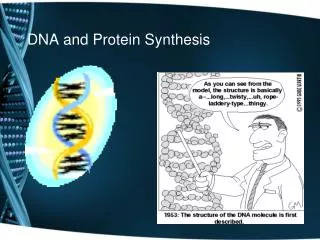
Non-Protein Nitrogen (NPN) Compounds (Urea, Creatinine & Uric Acid). Lab. 7. Introduction. Nitrogen containing compounds that are not proteins or polypeptides Total NPN can be tested by making a protein-free filtrate

E N D
Non-Protein Nitrogen (NPN) Compounds (Urea, Creatinine & Uric Acid) Lab. 7
Introduction • Nitrogen containing compounds that are not proteins or polypeptides • Total NPN can be tested by making a protein-free filtrate • Useful clinical information is obtained from individual components of NPN fraction
Urea • Highest concentration of NPN in blood (45%) • Major excretory product of protein metabolism • Urea is synthesized in the liver, from CO2 and ammonia, as the final product of amino acid catabolism. • It is freely filtered at the glomerulus, though 40% is passively reabsorbed by the proximal tubules.
Urea • Blood urea levels can vary proportionately with: • the protein content of the diet, • rate of protein catabolism during tissue breakdown, • and liver function. • Because of its metabolism, urea is a nonspecific indicator of renal function. • However, in healthy persons with an average diet, the blood urea level is a very sensitive indicator of renal function. • Blood urea levels are usually elevated before significant changes in creatinine levels have occurred. • Monitoring blood urea levels is very useful when one is following the course of renal disease.
Clinical Significance • States associated with elevated levels of urea in blood are referred to as uremia or azotemia. • Causes of urea plasma elevations: • Prerenal • Renal • and postrenal • Parallel determination of urea and creatinine is performed to differentiate between pre-renal and post-renal azotemia.
Specimen • Serum and heparinized plasma can be used for the urease/GLDH methods. • Fluoride will inhibit the urease reaction; therefore methods employing urease cannot use serum preserved with fluoride. • Ammonium heparin also cannot be used as an anticoagulant for urease methods. • Stability in serum or plasma: • 7 days at 4–8°C • 1 year at -20°C • Because of urea’s susceptibility to bacterial degradation, serum and urine samples should be kept at 4° to 8° C until analysis.
Specimen • One can analyze urine after a 1:20 to 1:50 or 1:100 sample dilution, depending on the method and instrument employed. • One can also preserve urine samples by maintaining the pH at less than 4. • Stability in urine: • 7 days at 4–8°C • 1 month at -20°C • Discard contaminated specimens.
Urease/GLDH Method • The method is optimized for 2-point kinetic measurement. • Decrease in absorbance at 340 nm is proportional to concentration of urea
Creatinine • Creatinine is a non-protein nitrogen waste product formed in muscle. Creatine Phosphate – phosphoric acid = Creatinine Creatine – water = Creatinine • Filtered by kidney and excreted in the urine • Creatinine filters easily into the glomerular filtrate and is not reabsorbed by the tubule. • The amount excreted daily is a function of muscle mass and is not affected too much by diet
Clinical Significance • Elevated Creatinine is found with abnormal renal function (i.e. GFR) • Therefore, any condition that reduces the glomerular filtration rate will result in: • a lessened excretion from the body, • with a consequent rise in the concentration of creatinine in the blood. • The serum creatinine is a better indicator of renal function than either that of BUN or uric acid
Clinical Significance • For renal transplant patients, an increase in serum creatinine of 2 mg/L has been used as a criterion of establishing rejection. • In other persons a change in creatinine of 2 mg/L would represent a 20% loss in renal function.
Specimen • One can analyze serum, plasma, or diluted urine. • The common anticoagulants (fluoride and heparin) do not cause interference, though heparin, which can be formulated as the ammonium salt, must be avoided in enzymatic methods that measure ammonia production. • Storage • 7 days at 4-25oC • At least 3 months at -20oC
Specimen • Urine should be diluted 1:100 • Bacterial contamination has been found to falsely lower creatinine values measured using the Jaffé reaction. • The mechanism of this interference appears to be bacterial production of a substance that retards the rate of the Jaffé reaction.
Jaffe Method Creatinine + Sodium PicrateCreatininr-picrate complex ( Yellow-orange) • The difference in absorbance at fixed times during conversion is proportional to the concentration of creatinine in the sample
Creatinine Clearance • Creatinine clearance is used to estimate the glomerular filtration rate (GFR). • Creatinine is chosen because it is freely filtered at the glomerulus and is not reabsorbed by the tubules. • However, a small amount of the creatinine (about 5%) in the final urine of healthy persons is derived from tubular secretion. • To do the test, one needs a precisely timed urine collection and a blood sample taken during the collection period.
Creatinine Clearance • Best results are obtained from a 24-h urine collection. • The test is initiated by having patients empty their bladder at the beginning of the timed period. • Urine is collected throughout the period, the bladder is again emptied at the end of the time period.
Creatinine Clearance • Creatinine determinations are performed on both samples. The creatinine clearance is calculated from the following formula: Creatinine clearance (mL/min)= (U x V)/P x 1.73/S • Where: • U is urinary creatinine (mg/L), • V is volume of urine (mL/min), • P is plasma creatinine (mg/L), • S is the calculated surface area of the patient, • and 1.73 is the surface area (m2) of a standard 70 kg person.
Creatinine Clearance • The range of creatinine clearance in healthy persons corrected to a surface area of 1.73 m2 is 90 to 120 mL/min. • At low filtration rates, the creatinine clearance does not parallel true glomerular filtration rate because a relatively large portion of the urine creatinine is secreted rather than filtered.
Parallel determination of urea and creatinine is performed to differentiate between pre-renal and post-renal azotemia. • Pre-renal azotemia, caused by e.g. dehydration, increased protein catabolism or decreased renal perfusion, leads to increased urea levels,while creatinine values remain within the reference range. • In post-renal azotemias, caused by the obstruction of the urinary tract, both urea and creatinine levels rise, but creatinine in a smaller extent.
Uric Acid • Uric acid is formed from the breakdown of nucleic acids and is an end product of purine metabolism. • Uric acid is transported by the plasma from the liver to the kidney, where it is filtered and where about 70% is excreted. • The remainder of uric acid is excreted into the GI tract.
Clinical Significance • Disease states with increased plasma uric acid • Gout • Increased catabolism of nucleic acids • And renal disease • In Gout increased serum levels of uric acid lead to formation of monosodium urate crystals around the joints. • Uric acid test is useful to assess for gout and to monitor patients with renal failure
Clinical Significance • To monitor if uric acid levels are too high after chemotherapy or radiation. • Hypouricemia is seldom observed and associated with rare hereditary metabolic disorders.
Specimen • Serum or plasma may be used • Stability in serum / plasma: • 6 months at -20°C • 7 days at 4-8°C • 3 days at 20-25°C
Enzymatic Colorimetric • Uric acid is oxidized to allantoin by uricase. • The generated hydrogen peroxide reacts with 4-aminophenazone/ESPT to quinoneimine. Aminophenazone + DHBS