TITLE: “SUBARACHNOID HAEMORRHAGE”
690 likes | 1.62k Vues
TITLE: “SUBARACHNOID HAEMORRHAGE”. Group Members : (1) STN Minsuin ak Akong (2) STN Siti Niza (3) STN Siatny ak Bujang (4) STN Sim Jia Ming

TITLE: “SUBARACHNOID HAEMORRHAGE”
E N D
Presentation Transcript
TITLE: “SUBARACHNOID HAEMORRHAGE” Group Members: (1) STN MinsuinakAkong (2) STN SitiNiza (3) STN SiatnyakBujang (4) STN SimJia Ming (5) STN Priscilla DayangNgilo (6) STN Monica Eemas (7) STN MistikaRinaiBaru (8) STN Nelson UjaiakSinggon
CONTENTS • Objective • Personal Data • Chief Complaint • History • Definition of SAH • Pathophysiology • Sign & symptom • Assessment -Parameter -Laboratory investigation -Radiology investigation 9. Management -Medical management -Surgical management 10. Nursing Care 11. Health education 12. Summary
Objective … • To improve knowledge on neurological topic • To improve understanding on pathophysiology of SAH • To relate the indication of medication to patient condition • To give opportunity to relate theory to practice regarding nursing care of the patient
Personal Data Name: Mr. L Medical Registration Number: 333273 Age: 49 yrs old Sex: Male Occupation: Mechanic Date Of Admission: 15/01/2013 Diagnosis: Subarachnoid haemorrhage
Chief Complaint • Patient having sudden onset of severe headache in the afternoon followed by comatose state at 5.00p.m on 14/1/13. • No history of fall and emotional disturbance. • Subsequently admitted to Sibu Hospital and he was intubated and ventilated. • Doctor(Sibu Hospital) was queried patient has aneurysm.
Family request to transferred to NORMAH to have further treatment and management. • Patient was transferred by helicopter on 15/01/2013 @ 1.30 a.m. • Admitted to ICU for further treatment and management.
History • Medical history: • Unknown medical problem • Surgical history: • NIL • Social history: • Married with 3 children • Alcoholic- since patient was 14 years old. • Non-smoker
What is aneurysm? • A balloon-like bulge or weakening of an artery wall that ruptures, releasing blood into the subarachnoid space around the brain.
When red blood cells break down, toxins can cause the walls of arteries nearby to contract and spasm. The larger the SAH, the higher the risk of vasospasm. • A ruptured aneurysm releases blood into the subarachnoid space (left).
Subarachnoid haemorrhage • Subarachnoid haemorrhage isbleeding into the subarachnoid space-the area between the arachnoid membrane and the pia mater surrounding the brain. This may occur spontaneously, usually from a ruptured cerebral aneurysm.
The difference between a normal brain and a brain that has SAH
Not known medical illness Aneurysms typically form in the bifurcations of the large vessels that make up the circle of Willis.
Aneurysm leaking Leading to blood extravasation into the subarachnoid space.
Subarachnoid hemorrhage Query rogressivedecrease in cerebral blood flow occurs, result from a sudden massive increase in intracranial pressure (ICP)
Assessment • Vital signs: • On admission- 15/1/13, • B/P: 220/90mmHg • PR: 85 bpm • RR: 15 bpm • Temp. : 37.5°C • SPO₂: 100% ventilated with 40% O₂ • An invasive line was inserted • Arterial line • Central Venous Catheter (CVC) • Intracranial Pressure (ICP)- 16-20mmHg • Cerebral Perfusion Pressure (CPP)- 75mmHg
Vital signs during hospitalization in ICU (16/1/13-24/1/13): (Ranging) • B/P: 150-160/ 60-80mmHg • PR: 83- 90bpm • RR: 16- 21bpm • Temp.: 37.5°C- 39.4°C • SPO₂: 96%-100% ventilated with 40% O₂ • On 15/1/13 @3p.m – 130/50 (IV Noradrenaline was given as per Dr. order) *Off on 16/1/13 @9a.m
GCS (Glasgow Coma Scale): • Patient’s GCS was at the range of 5/15 – 11/15 during hospitalization in ICU. • On the GCS motor assessment, patient’s Lt. sided was totally not response to our pain stimulisation. However, his Rt. sided was slightly weak and move flexion when response to pain stimulisation. • On the eye assessment, patient looked drowsy, only open eyes to call and not obey command.
Both pupil size was at the range of 2-4mm and reactive to light.
All invasive line and ventilator was off on 22/1/13. Parameter was stable. • No more ICP and CPP monitoring. • Patient was transferred out to Serapi ward on 24/1/13 @9.45 a.m with tracheostomyinsitu. • Vital signs was stable before transferred to Serapi ward except patient was still having fever (37°C-38°C). • On trachy ventilated with 1L of O₂
Diagnostic Procedure • Lab test • Radiological
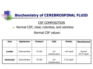
LaboratoryTest 1.Heamatology
Laboratory Test 1.Heamatology
Renal Function TestObjectives : To evaluate how well the kidney are working.Types of test carried out :-
Liver Function TestObjectives : To detect inflammation and damage to the liver and to evaluate how well liver working. Types of test carried out :-
Lipid ProfileObjectives : to evaluate the abnormalities in lipid level in the blood. Types of test carried out :-
Culture and sensitivity(C&S)Objective :-A culture is done to find out what kind of organism (usually a bacteria) is causing an illness or infection. -A sensitivity test checks to see what kind of medicine, such as anantibiotic,will work best to treat the illness or infection. Date : 15/1/13 Specimen : Blood Test Ordered : Urin , spuctum,CSF Result : No growth after 48hour Date: 18/1/13 (repeat) Specimen: Sputum Test Ordered : Blood ,urine , CSF Result : Klebsiellapneumoniae ( Tazocin ) Date : 23/1/13 (repeat) Specimen : Sputum taken at ETT Test Ordered : Blood ,urine , CSF Result : Acinetobacterbaumannii
Radiological investigation • CXR • MSCT • Cerebral angiogram
Chest X-RayObjective: Used to screen ,diagnose and evaluate changes in respiratory system. Date: 15/1/13 Result: -Left mid zone consolidation. (alveoli stick to each other)
Multislice Computed Tomography ScanObjective: To visualize the heart anatomy, coronary circulation and great blood vessel. Date : 15/1/13 Result :- -Right frontal bleed with massive subarachnoid extension. Date: 16/1/13 Result:- -No hydrocephalus or increase in bleeding noted. -The brain does not show any mass effect.
Operation: Cerebral Angiogram Date: 15/1/13@2.50pm Definition of cerebral angio? Cerebral angiography is a form of angiography which provides images of blood vessels in and around the brain, thereby allowing detection of abnormalities such as arteriovenous malformations and aneurysms. Result Cerebral Angiogram:- Clinical problem- ? ACAA Conclusion:- -Anterior communicating artery aneurysm.(ACAA)
Medical Management • G:\ICU CASE STUDY - medical.docx
Operation : Tracheostomy Date :19/1/13 Definition of Tracheostomy? -A tracheostomy is a surgical procedure to create an opening through the neck into the trachea (windpipe). A tube is usually placed through this opening to provide an airway and to remove secretions from the lungs. This tube is called a tracheostomy tube or trach tube.
Operation:Left Craniotomy & Clipping of aneurysm Date: 21/1/13 Definition of craniotomy? A craniotomy is the surgical removal of part of the bone from the skull to expose the brain. Specialized tools are used to remove the section of bone called the bone flap. The bone flap is temporarily removed, then replaced after the brain surgery has been performed.
Definition of aneurysm clipping? The goal of surgical clipping is to isolate an aneurysm from the normal circulation without blocking off any small perforating arteries nearby. A small clip is placed across the base, or neck, of the aneurysm. Blades of the clip remain tightly closed until pressure is applied to open the blades.