Download
1 / 53
530 likes | 1.48k Vues
MICROORGANISMS OF THE SKIN AND MUCOUS MEMBRANES. Microbial flora of the skin and mucous membranes : Resident flora, usually commensal microorganisms 2. Pathogenic microorganisms. Role of the resident flora. Role of resident flora of the skin and mucous membranes:

E N D
MICROORGANISMS OF THE SKIN AND MUCOUS MEMBRANES Microbial flora of the skin and mucous membranes : Resident flora, usually commensal microorganisms 2. Pathogenic microorganisms
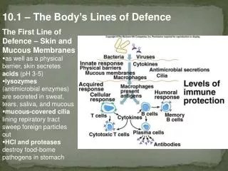
Role of the resident flora Role of resident flora of the skin and mucous membranes: To prevent colonization by pathogens and possible disease through bacterial interference. The mechanisms of bacterial interference : 1. competition for receptors or binding site on host cells 2. competition for nutrients 3. mutual inhibition by metabolic or toxic products 4. mutual inhibition by antibiotic materials or bacteriocins
Role of the resident flora • Suppression of the normal flora creates a partial local void that tend to be filled by microorganisms from the environment or from other part of the body. • Such organisms behaves as opportunists and may be become pathogen
Role of the resident flora • Members of the normal flora may themselves produce disease under certain circumstances • These organisms are adapted to the noninvasive mode of life defined by the limitations of the environment • If forcefully remove the restrictions of that environment and introduced into the blood stream or tissues, these organisms may become pathogenic • Large numbers of Streptococcus viridans (normal flora of the upper respiratory tract ) introduced into the bloodstream (following tooth extraction or tonsillectomy), they may settle on deformed heart valve and produce infective endocarditis
Normal flora of the skin • The skin is particularly apt to contain transient microorganisms, because of its constant exposure to and contact with the environment • There is a constant and well-defined resident flora, modified in different anatomic area by secretions, proximity to mucous membranes (mouth, nose, perineal areas) , and habitual wearing of clothing • The factors that may be important in eliminating nonresident microorganism from the skin are the low pH, the fatty acid in sebaceous secretions, and the presence of lyzozyme
Normal flora of the skin • Neither profuse sweating nor washing and bathing can eliminate or significantly modify the normal resident flora • The number of superficial microorganisms may be diminished by vigorous daily scrubbing with soap, but the flora is rapidly replenished from sebaceous and sweat glands even when contact with other skin area or with the environment is completely excluded • Placement of an occlusive dressing on the skin tends to result in a large increase in the total microbial population and may also produce qualitative alterations in the flora
Normal flora of the skin • Anaerobic and aerobic bacteria often to join to form synergistic infections ( gangrene, necrotizing fasciitis, cellulitis) of skin and soft tissues • The bacteria are frequently part of the normal microbial flora • It is usually difficult to pinpoint one specific organisms as being responsible for the progressive lesion, since mixtures of organisms are usually involved
Normal flora of the skin • Staphylococcus epidermidis • Staphylococcus aureus ( in small numbers ) • Alpha-hemolytic and nonhemolytic Streptococcus • Micrococcus species • Peptostreptococcus species • Neisseriae species ( nonpathogenic ) • Propionibacterium species • Diphtheroids • Candida species ( small numbers ) • Acinetobacter species ( small numbers )
Genus Staphylococcus • Cells spherical, 0,5 – 1,5 um in diameter, occurring singly, in pair and irregular cluster • Gram positive, nonmotile, nonsporing • Facultative anaerobic, chemoorganotrophic,with both respiratory and fermentative metabolism • Colonies are usually opaque and may be white or cream and sometimes yellow to orange • Usually catalase positive, cytochromes present but usually oxidase negative • Nitrate often reduce to nitrite, susceptible to lysis by lysostaphin but not by lyzozyme • Usually grow with 10 % NaCl, the optimum temperature is 300-370C • Mainly associated with the skin and mucous membranes of warm-blooded vertebrates, but are often isolated from food products, dust and water • Some species produce extracellular toxin
Tabel 1. Laboratory test for differentiation of Staphylococcal sp.
Genus Streptococcus • Cells spherical or ovoid, 0,5 – 2,0 um in diameter, occurring in pairs or chains when grown in liquid media; they sometimes elongated in the axis to a lanceolate shape • Gram positive, nonmotile, nonsporing, some sp are encapsulated • Facultatively anaerobic, chemoorganotrophs, requiring nutritionally rich media for growth and sometime 5 % CO2 • Metabolism fermentative, producing mainly lactate but no gas • Catalase negative, commonly attack red blood cells, with either greenish discolorization ( alpha hemolysis ) or complete clearing (beta hemolysis ), growth is usually restricted to a temperature of 250 – 450 C (optimum 370C ) • Mainly inhabiting the mouth and upper respiratory tract
Genus Micrococcus • Cells spherical, 0,5 – 2,0 um in diameter, occurring in pairs, tetrads or irregular cluster, not in chains • Gram positive, seldom motile, nonsporing • Strictly aerobic; collonies usually pigmented in shades of yellow or red, usually grow on simple media • Chemoorganotrophs, with a respiratory metabolism, often producing little or no acid from carbohydrates, catalase positive and often oxidase positive, usually halotolerant, grow with 5% NaCl • Contain cytochromes and are resistant to lysostaphin • The optimum temperature is 250 – 370 C, occur primarily on mammalian skin and in soil but commonly are isolated from food products and the air
Genus Peptostreptococcus • Cells spherical, 0,5 – 1,2 um in diameter, and sometimes ovoid; arrangement is variable; in pair, tetrads, clumps, or chains. • Gram positive, nonmotile, nonsporing • Anaerobic, chemoorganotrophic and fermentative, requiring nutritionally rich media, metabolize peptone to mainly acetic acid; their attack on carbohydrates is usually weak or absent • Usually catalase negative, but weak or pseudocatalase reactions may occur; some members produce indole and reduce nitrate • The optimum temperature is 370 C • The genus is differentiated from Peptococcus mainly by its lower mol % G+C content of the DNA ( 27 – 45 ) • Obligate parasites of the mouth, mucous membranes, and intestinal tract of mammals, and may play a part in purulent infections
Genus Neisseria • Cocci are 0,6 1,0 um in diameter, occurring singly but more often in pairs with adjacent sides flattened; one species (N. elongata) is an exception and consists of short rods 0,5 um wide, often arranged as diplobacilli or in short chains • Division of the coccal species is in two planes at right angle to each other, sometimes resulting in tetrads • Capsules and fimbriae (pili) may be present; endospore are not present • Cells stain gram negative, but there is tendency to resist decolorization • Swimming motility does not occur, and flagella are absent • Aerobic; some species produce a greenish yellow carotenoid pigment
Genus Neisseria • Some species are nutritionally fastidious and hemolytic, optimum temperature is 350 – 370 C • Oxydase positive, catalase positive, except N. elongata; carbonic anhydrase is produced by all species; all species reduce nitrite except N. gonorrhoeae and N. canis. • Chemoorganotrophic, some species are saccharolytic • They are inhabitants of the mucous membranes of mammals • Some species are primary pathogens for humans
Genus Propionibacterium • Pleomorphic rods, 0,5-0,8x1-5 um, are often club shaped with one end rounded and the other tapered; some cells may be coccoid, bifid or branched, but they are not filamentous; cells occur singly, in pairs or short chains, in V or Y configurations, or in clumps with “chinese character” arrangement • Gram positive, non motile, nonsporing • Facultative anaerobes but have variable aerotolerance; most grow somewhat in air but better anaerobically, giving on blood agar colonies that are usually convex, semi opaque, glistening, and often pigmented in shades of cream to reddish • Chemoorganotrophic with complex nutritional requirement, have a metabolism fermentative, producing from glucose and some other carbohydrates large amounts of propionic acid , acetic acid and often small amounts of gas
Genus Propionibacterium • The optimum growth temperature is 300 – 370 C • Usually catalase positive; they are found mainly in cheese and dairy products and on human skin • Readily confused with some species of Corynebacterium or Clostridium
Genus Corynebacterium • Straight or slightly curved, slender rods have tapered or sometimes clubbed ends and are 0,3-0,8 x 1,5-8,8 um • Cells are usually arranged singly or in pairs, often in a V formation or in palisade of several parallel cells • Gram positive, though some cells stain unevenly, giving beaded appearance; metachromatic granules of polymethaphosphate are commonly formed within the cells • Nonmotile, nonsporing,not acid-fast • Facultative anaerobs, commonly requiring nutritionally rich media such serum or blood media, on which colonies are usually convex and semi opaque, with a mat surface • Chemoorganotrophs with fermentative metabolism, most species produce acid without gas from glucose and some other carbohydrates
Genus Corynebacterium • Catalase positive, often reduce nitrate and tellurite; rarely acidify lactose or raffinose or liquefy gelatin • Primarily obligate parasites of mucous membranes of skin of mammals; but occasionally they are found in other sources; some species are pathogenic for mammals
Normal flora of the mucous membranes Normal flora of the : • Conjunctiva • Mouth and upper respiratory tract • Intestinal tract • Urethra • Vagina
Normal flora of the conjunctiva The predominant organisms of the conjunctiva are : • Diphtheroids • Staphylococcus epidermidis • Streptococcus (nonhemolyticus) • Neisseriae • Moraxella sp The conjunctival flora is normally held in check by the flow of tears; which contain antibacterial lyzozyme
Normal flora of the mouth and upper respiratory tract The flora of the nose consist of : - corynebacteria - Staphylococcus epidermidis - Staphylococcus aureus - streptococci
Normal flora of the mouth and upper respiratory tract • The mucous membranes of the mouth and pharynx are often sterile at birth but may be contaminated by passage through birth canal • Within 4 – 12 hours after birth, viridans streptococci become established as the most prominent members of the resident flora and remain so for life. They probably originate in the respiratory tracts of the mother and attendants. • Early in life, aerobic and anaerobic staphylococci, gram negative diplococci ( Neisseriae, Moraxella catarrhalis ), diphtheroids, and occasional lactobacilli are added
Normal flora of the mouth and upper respiratory tract • When teeth begin to erupt, the anaerobic spirochetes, Prevotella sp (especially P. melaninogenica ), Fusobacterium sp, Rothia sp, and Capnocytophaga sp established themselves, along with some anaerobic vibrios and lactobacilli • Actinomyces sp are normally present in tonsillar tissue and on the gingivae in adults, and various protozoa may also present. Yeast (Candida sp) occur in the mouth • In the pharynx and trachea, a similar flora established itself, whereas few bacteria are found in normal bronchi. Small bronchi and alveoli are normally sterile
Normal flora of the mouth and upper respiratory tract • The predominant organisms in the upper respiratory tract, particularly in the pharynx : • Nonhemolytic streptococcus • Alpha hemolytic streptococcus • Neisseriae • Staphylococci • Diphtheroid • Haeomophili • Pneumococci • Mycoplasma • Prevotella
Normal flora of the mouth and upper respiratory tract • Infections of the mouth and respiratory tract are usually caused by mixed oronasal flora, including anaerobes • Periodontal infections, perioral abscess, sinusitis and mastoiditis may involved predominantly Prevotella melaninogenica, Fusobacteria and Peptostreptococci • Aspiration of saliva (containing up to 102 of these organisms and aerobes) may results in necrotizing pneumonia, lung abscess and empyema
Normal flora of the intestinal tract • At birth the intestine is sterile, but organisms are soon introduced with food. In breast-fed children, the intestine contain large numbers of lactic acid streptococci and lactobacilli. These aerobic and anaerobic, gram positive, nonmotile organisms (e.g. Bifidobacterium species) produced acid from carbohydrates and tolerate pH 5.0 • In bottle-fed children, a more mixed flora exist in the bowel, and lactobacilli are less prominent. As food habits develop toward the adult pattern, the bowel flora changes • Diet has a marked influence on the relative composition of the intestinal fecal flora • Bowels of newborns in intensive care nurseries tend to be colonized by Enterobacteriaceae, e.g. Klebsiella, Citrobacter, Enterobacter
Normal flora of the intestinal tract • In normal adults, the esophagus contains microorganisms arriving with saliva and food. • The stomach’s acidity keep the number of microorganisms at minimum (103 – 105 /gr content ) unless obstruction at the pylorus favors the proliferation of gram positive cocci and bacilli. The normal acid pH of the stomach markedly protects against infection with some enteric pathogens, e.g. cholera • Administration of cimetidine for peptic ulcer leads to great increase in microbial flora of the stomach, including many organisms usually prevalent in feces
Normal flora of the intestinal tract • As the pH of intestinal content become alkaline, the resident flora gradually increases. In the adult duodenum, there are 103 -106 bacteria per gram of content; in the jejunum and ileum, 105-108 bacteria/gr; and in the cecum and transverse colon, 108-1010 bacteria/gr • In the upper intestine, lactobacilli and enterococci predominate, but in the lower ileum and cecum, the flora is fecal • In the sigmoid colon and rectum, there are about 1011 bacteria/gr of content, constituting 10 – 30% of the fecal mass
Normal flora of the intestinal tract • Anaerobes outnumber facultative organisms by 1000-fold. In diarrhea the bacterial content may diminish greatly, whereas in intestinal stasis the count rises • In the normal adult colon, 96 – 99% of the resident bacterial flora consists of anaerobes : • Bacteroides sp, especially B. fragilis • Fusobacterium sp • Anaerobic lactobacilli, e.g. bifidobacteria • Clostridia ( C.perfringens, 103 -105/gr) • Anaerobic gram positive cocci (Peptostreptococcus sp)
Normal flora of the intestinal tract • Only 1 – 4% are facultative aerobes : • Gram negative coliform bacteria • Enterococci • Small number of protei, pseudomonads, lactobacilli, candidae • More than 100 distinct types of organisms, which can be cultured routinely in the laboratory, occur regularly in the normal fecal flora • There probably are more than 500 sp of bacteria in the colon including many that are likely unidentified. Minor trauma(e.g. sigmoidoscopy, barium enema) may induce transient bacteremia in about 10% of procedures
Normal flora of the intestinal tract Intestinal bacteria are important in : • Synthesis of vitamin K • Conversion of bile pigments and bile acids • Absorption of nutrients and breakdown products • Antagonism to microbial pathogens • The intestinal flora produces ammonia and other breakdown products that are absorbed and can contribute to hepatic coma • Among aerobic coliform bacteria, only few serotypes persist in the colon for prolonged periods, and most serotypes of Escherichia coli are present only over period of a few days
Normal flora of the intestinal tract • Antimicrobial drugs taken orally can, in human, temporarily suppress the drug susceptible component of the fecal flora • This is commonly done by preoperative oral administration of insoluble drug. For example, neomycin plus erythromycin can in 1 – 2 days suppress part of the bowel flora, especially aerobes • Metronidazole accomplishes that for anaerob. If lower bowel surgery is performed when the counts are at their lowest, some protection against infection by accidental spill can be achieved • However, soon thereafter the counts of fecal flora rise again to normal or higher than normal levels, principally of organisms selected out because to relative resistance to the drug employed
Normal flora of the intestinal tract • The drug susceptible microorganisms are replace by drug resistant ones, particularly staphylococci, enterobacters, enterococci, protei, pseudomonads, Clostridium difficile and yeast • The feeding of large quantities of Lactobacillus acidophilus may results in the temporary establishment of this organisms in the gut and the concomitant partial suppression of other gut microflora • The anaerobic flora of the colon, including B. fragilis, clostridia and peptostreptococci , play a main role in abscess formation originating in perforation of the bowel. • Prevotella bivia, P. disiens are important in the abscesses of the pelvis originating in the female genital organ • These sp are penicillin-resistant
Normal flora of the urethra • The anterior urethra of both sexes contains small numbers of the same types of organisms found on the skin and perineum • This organism regularly appear in normal voided urine in numbers of 102 – 104/mL
Normal flora of the vagina • Soon after birth, aerobic lactobacilli appear in the vagina and persist as long as the pH remains acids (several weeks) • When pH become neutral (remaining so until puberty), a mixed flora of cocci and bacilli is present • At puberty, aerobic and anaerobic lactobacilli reappear in large numbers and contribute to the maintenance of acid pH through the production of acid from carbohydrates, particularly glycogen • This appears to be an important mechanisms in preventing the establishment of other, possibly harmful microorganism in the vagina
Normal flora of the vagina • If lactobacilli are suppressed by the administration of antimicrobial drugs, yeast or various bacteria increase in numbers and cause irritation and inflammation • After menopause, lactobacilli again diminish in number and mixed flora returns. The normal vaginal flora includes group B streptococci in as many as 25% of women of childbearing age. • During the birth process, a baby can acquire group B streptococci, which subsequently may cause neonatal sepsis and meningitis • The normal vaginal vaginal flora often includes also alpha hemolytic streptococci, anaerobic streptococci( peptostreptococci), Prevotella sp , clostridia, Gardnerella vaginalis, Ureaplasma urealyticum, and sometimes listeria or Mobiluncus sp
Normal flora of the vagina • The cervical mucus has antibacterial activity and contain lyzozyme. In some women, the vaginal introitus contain a heavy flora resembling that the perineum and perianal area • This may be a predisposing factor in recurrent urinary tract infections • Vaginal organisms present at time of delivery may infect the newborn (e.g. group B streptococci )
Skin and soft tissue infections:Staphylococcus aureus • Attributes of pathogenicity of S. aureus : • Coagulase enhance fibrin deposition and abscess formation. There is also clumping factor that coats the cells with fibrin • Cytolytic toxin (alpha, beta, gamma, delta and leukocidin ) are all hemolytic (except leukosidin) and destroy cellular membranes • TSST-1 formerly termed enterotoxin F, is a superantigen and toxin produced under certain environmental conditions, most commonly associated with tampon use and surgical packing. • TSST-1 reduces liver clearance of endogenous endotoxin • Exfoliatins produced by phage group II S. aureus cause surface layer of the skin to separate (probably through disruption of intracellular junctions) leading to desquamation
Attributes pathogenicity of S. aureus • Protein A ( a surface protein ) is anti phagocytic (binding to the Fc portion of antibody, making it unavailable to attach to phagocytes • Teichoic acids aids in attachment and stimulate the inflamatory response when complexed with peptidoglycan
Clinical disease • Skin infections include impetigo (often bullous), folliculitis of the bearded region, boils (furuncles), carbuncles (more extensive), styes, and surgical wound, burn, or traumatic-lession infections • Scalded skin syndrome, with its characteristic bullae and desquamation of body surfaces, occur most commonly in children younger than 5 years old, sometimes with fairly minor infections but circulating exfoliatins
Skin and soft tissue infections :Clostridium perfringens General characteristic of Cl. perfringens : • Anaerobic, spore-forming, large gram positive rod • Spore can be central or subterminal and relatively heat resistant • Has soil as natural habitat; contamination can occur in home-canned goods, smoked fish, and honey • Has germination of spores and emergence of vegetative cells as being necessary for toxin production
Attributes of pathogenicity of Cl. perfringens • Produced alpha toxin, a potent lecithinase that damage cellular membranes • Produce 11 other toxins or enzymes that damage eukaryotic cells • Produces an enterotoxin associated with food poisoning
Clinical disease Cl. perfringens cause two types of infections : • Soft tissue wound infections following severe trauma; organisms elaborate toxins and enzymes to produce gas, edema, and impaired of circulation; vascular destruction and lactic acid accumulation lower the redox potential, with two consequences : 1. anaerobic cellulitis, causing destruction of traumatized only 2. myonecrosis (gas gangrene) or destruction of traumatized tissue and surrounding healthy tissue
Skin and soft tissue infections :Pseudomonas aeruginosa General characteristic of P. aeruginosa : • A small, polarly flagellated, gram negative rod with pili • A non fermentative, oxydase positive bacterium • A ubiquitous environmental organism found in water and soil and widely distributed on plants • It can grow in both distilled water or tap water overnight to large number • Often produces pigments that may be clinically useful, such as fluorescein ( pyoverdin ) , a greenish fluorescent pigment, and pyocyanin, a blue-green pigment • Blue-green pus is a classic sign of P. aeruginosa infection
Attributes pathogenicity of P. aeruginosa Invasive factor includes : • Pili, which adhere • A polysaccharide slime layer, which increases adherence to tissue, making them less susceptible to phagocytosis Virulence factor includes : • Exotoxin A, an ADP ribose transferase similar to diphtheria toxin, which inactivates the tRNA elongation factor (EF 2), halt protein synthesis, and causes liver necrosis • Exoenzyme S , an ADP ribose transferase capable of inhibiting eukaryotic protein synthesis • Lipopolysaccharide • Phospolipase C, which damages membranes causing tissue damage • Elastase and other proteolytic enzyme
Clinical disease Cellulitis : • Occur in patient with burns, wound, or neutropenia; may be highly necrotic; indicated by blue-green pus and grape-like sweet odor Septicemia : • Results from hematogenous spread of infection from local lesion or gastrointestinal tract and causes gram negative shock • May result in a distinctive lesion, ecthyma gangrenosum, when dermal veins and tissue are invaded. These lesions become necrotic
Diagnosis • Most commonly made by clinical suspicion (grape-like odor, blue-green pus or ecthyma gangrenosum) and confirm by culture • Shows beta hemolysis on blood agar, with pigment production • Shows nonfermentation on macConkey agar, blue-green pigment, grape like odor, and oxydase positivity
Skin and soft tissue infections :Streptococcus pyogenes General characteristic of S. pyogenes : • Occur as single,paired, or chained gram positive cocci, depending on the environment, facultative anaerobe, attaches to epithelial surface via lipoteichoic acid portion of fimbriae (pili) • Classification : • Classified as group A of the 21 Lancefield group of streptococci, which are distinguished serologically by slight differences in specific cell wall carbohydrates • Contains group A- specific carbohydrate and several antigenic protein (M,T and R antigen) in the cell wall • Subdivided into more than 80 types base on antigenic differences in the M protein
Skin and soft tissue infections :Streptococcus pyogenes • General characteristics of S. pyogenes : • Sensitive to bacitracin, catalase negative, rarely become resistant to penicillin