CNS
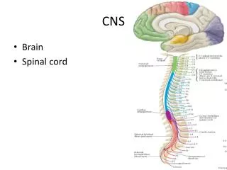
CNS. Brain and spinal cord Brain = two cerebral hemispheres, brainstem, cerebellum, Diencephalon = thalamus/hypothalamus/pituitary Spinal cord = ascending and descending axons, interneurons, autonomic neurons, sensory neurons, motor neurons.


CNS
E N D
Presentation Transcript
CNS Brain and spinal cord Brain = two cerebral hemispheres, brainstem, cerebellum, Diencephalon = thalamus/hypothalamus/pituitary Spinal cord = ascending and descending axons, interneurons, autonomic neurons, sensory neurons, motor neurons Cerebral hemispheres divided into lobes; connected by a layer of axons called the corpus callosum; takes care of collection and integration of sensory input, regulation of mood and behavior, and higher order thought processes Brainstem divided based on development into areas called the midbrain, the pons and the medulla; regulates basic functions such as breathing, heart rate, and digestion
Cerebrum Association areas = integration and direction of voluntary behaviors Fields = areas where sensory information comes in and is integrated into perception
Limbic system • Amygdala – emotion and memory • NMDA receptors • Hippocampus – learning and memory • NMDA receptors • GABA receptors • Basal ganglia (corpus striatum) – control of movement • GABA receptors
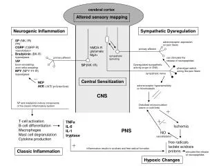
The NMDA receptor complex. Activation (i.e., excitation) occurs when either glutamate (Glu) or N-methyl-d-aspartate (NMDA) and glycine (Gly) bind to the receptor molecule. A channel within the receptor complex enables molecules to cross the cell membrane. Magnesium (Mg) blocks this channel. When Mg is removed from the channel and the receptor is activated, calcium (Ca++) and sodium (Na+) ions enter the cell and potassium ions (K+) leave. These cells can stay depolarized for long periods of time (up to days in some receptors). This is the basis of memory formation and is termed long term potentiation.
Diencephalon • Thalamus – relay station for sensory information to limbic system, cortex, cerebellum • GABA, serotonin, and dopamine receptors and neurons • Hypothalamus – centers for behavioral drives, biological clock, key in homeostasis mechanisms
Cerebellum • Interprets sensory information and produces appropriate coordinated movement • Norepinephrine is major neurotransmitter
Brainstem • Divided into three main parts: medulla oblongata, pons, mesencephalon (midbrain) • Cranial nerves emerge from this area; sensory and motor information to/from the head and neck, as well as the vagus nerve that innervates and receives information from many internal organs • Medulla = cross-over of information; control centers for blood pressure, breathing, swallowing, vomiting • Pons – relay station for information going between cerebellum and cerebrum • Mesencephalon – controls eye movement; relays for auditory and visual cortex Reticular formation runs through the brainstem – arousal/sleep, muscle tone, modulation of pain
Function in cerebral hemispheres is not identical; “right brain-left brain” • Hemispheres communicate with one another through a large set of myelinated axons called the corpus callousm What generalization can you make about right brain function? Left brain function? Which side would be “dominant” in right handed people?
Types of Sensory Receptors: • Chemoreceptors • Examples: • Mechanoreceptors • Examples: • Thermoreceptors • Examples: • Photoreceptors • Examples: • Nociceptors • Examples: Muscle tension/length, blood pressure, pH/O2 content of blood, pH of CSF, lung inflation, osmolarity of body fluids, blood glucose, distention of gut
Somatosensory neurons • Receptors can be free nerve endings or more complex receptors covering the nerve endings. • Axons can be either myelinated or unmyelinated • Heat, cold, pain, touch, pressure, proprioception • Conscious perception of information
Special Senses • Highly specialized receptors; some are neural in origin and morphology, many are non-neural in origin and morphology • Non neural = taste buds (epithelial cells), photoreceptors • Neural = smell
Characteristics of Sensory Neurons • Receptive fields; convergence results in a larger perceived receptive field • Stimuli converted into graded potentials or change in membrane potential • Threshold is the minimum stimulus required to activate a receptor • Coding and processing of stimuli allows us to determine the stimulus type, intensity, location, and duration • Type determined by the cortex in response to where the input comes from; 1:1 association between type of receptor and sensation is called labeled line coding • Location determined by which group of neurons in the cortex is activated; topographical organization of the sensory areas of the cortex; lateral inhibition is also used • Intensity of stimulus – population coding and frequency coding • Duration of stimulus – duration of action potentials; some neurons turn off after a certain amount of time (adaptation) • Tonic receptors – slow to adapt • Phasic receptors – adapt rapidly
Autonomic Nervous System • Often work in opposition • Cooperate to fine-tune homeostasis • Both may fire tonically; dominant system produces effect • Regulated by the brain; hypothalamus, pons and medulla • Can also be regulated by spinal reflexes; no higher order input • Pathways both consist of a two neuron system Preganglionic neuron autonomic ganglion postganglionic neuron target from CNS outside CNS
Parasympathetic • Sometimes called the “cranio-sacral division • Long preganglionic neurons; short postganglionic neurons (often in the target organ) • Preganglionic neurons secrete Ach on to nicotinic receptors • Postganglionic neurons secrete Ach on to muscarinic receptors • Target tissues are smooth muscle, cardiac muscle, exocrine glands, brown fat
Brancial motor(special visceral efferent) Supplies the voluntary muscles of the pharynx and most of the larynx, as well as one extrinsic muscle of the tongue. Visceral motor(general visceral efferent) Parasympathetic innervation of the smooth muscle and glands of the pharynx, larynx, and viscera of the thorax and abdomen. Visceral sensory(general visceral afferent) Provides visceral sensory information from the larynx, esophagus, trachea, and abdominal and thoracic viscera, as well as the stretch receptors of the aortic arch and chemoreceptors of the aortic bodies . General sensory(general somatic afferent) Provides general sensory information from the skin of the back of the ear and external auditory meatus, parts of the external surface of the tympanic membrane, and the pharynx. Special sensory(special afferent) A very minor component of CN X. Provides taste sensation from the epiglottic region. This component will not be discussed further.
Sympathetic • Sometimes called the “thoraco-lumbar” division • Short preganglionic neurons; long postganglionic neurons; ganglia are called the chain ganglia • Preganglionic neurons secrete Ach onto nicotinic receptors • Postganglionic neurons secrete NE on to a or b receptors • Target tissues are smooth muscle, cardiac muscle, endocrine glands, brown fat
B1 found on heart muscle and in certain cells of the kidney B2 found in certain blood vessels, smooth muscle of airways; found where sympathetic neurons ARE NOT A1 receptors are found most commonly in sympathetic target tissues A2 receptors are found in the GI tract and pancreas (relaxation)
Sympathetic nerve endings also activate the release of NE and E from the adrenal medulla Enhances effects of NE from sympathetic nerve endings Adds the effects of E to the overall arousal (“fight or flight”) pattern