Insights into Cortical Areas for Visual Processing of the Human Body Using fMRI
This presentation explores the cortical area selectively involved in visual processing of human body parts, based on fMRI studies. It covers fundamental concepts in functional neuroimaging and provides an overview of data analysis methodologies using Statistical Parametric Mapping (SPM). Key findings reveal functional segregation and integration in cortical areas, emphasizing physiological responses to sensorimotor and cognitive challenges. The presentation also discusses the intricacies of fMRI technology, including Blood Oxygenation Level Dependent (BOLD) contrast, spatial normalization, and noise reduction techniques critical for accurate data interpretation.

Insights into Cortical Areas for Visual Processing of the Human Body Using fMRI
E N D
Presentation Transcript
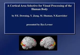
A Cortical Area Selective for Visual Processing of the Human Bodyby P.E. Downing, Y. Jiang, M. Shuman, N.Kanwisherpresented by Ilya Levner
Outline • Functional neuro-imaging • Issues • fMRI basics • Data analysis with SPM • Experiment Summary • Conclusions
Integration vs Segregation • Are physiological changes elicited by sensorimotor or cognitive challenges best characterized by • Functional Segregation • the activation of functionally segregated areas • Functional Integration • Anatomically distributed, but functionally integrated system
fMRI Basics • Based on BOLD Contrast • Blood Oxygenation Level Dependent contrast. • Deoxyhemoglobin is more paramagnetic (magnetizes easier) than oxyhemoglobin • So deoxyhemoglobin is the contrasting agent revealing cortical areas metabolizing oxygen at highter rates (presumably).
fMRI (Cont) • Small contrast difference oxygenated and deoxygenated blood. • 1.5T fMRI produce image intensity changes of no more than 2-4%. • 2T fMRI produce image intensity changes of less than 15%. • 4T fMRI has produced contrast changes of up to 30%
Statistical parametric map (SPM) Data Analysis Overview Design matrix Time-series data Kernel Realignment Smoothing General linear model Gaussian field theory Statistical inference Normalisation p <0.05 Template Parameter estimates
Reasons for Motion Correction The Steps in Motion Correction • registration - i.e. determining the 6 parameters that describe the rigid body transformation between each image and a reference image. • transformation - i.e. re-sampling each image according to the determined transformation parameters. • Subjects will always move in the scanner. • movement may be related to the tasks performed. • The sensitivity of the analysis is determined by the amount of residual noise in the image series, so movement that is unrelated to the task will add to this noise and reduce the sensitivity.
Spatial Normalisation Spatially normalised Original image • Enable reporting of activations as co-ordinates within a known standard space • e.g. the space described by Talairach & Tournoux • Inter-subject averaging • extrapolate findings to the population as a whole • increase activation signal above that obtained from single subject Spatial Normalisation Template image Deformation field
Smoothing • Why Smooth? • Potentially increase signal to noise. • Inter-subject averaging. • Increase validity of SPM. • In SPM, smoothing is a convolution with a Gaussian kernel. • Kernel defined in terms of FWHM (full width at half maximum). Before convolution After convolution Gaussian smoothing kernel
Statistical Parametric Maps (SPM) • Construction of statistical processes to test hypotheses about regionally specific effects. (functional segregation hypothesis) • Analysis of each voxel done using any standard (univariate) statistical test. • Voxel is a 3-D volumetric pixel.
Voxel by voxel statistics… model specification parameter estimation hypothesis statistic statistic image or SPM f MRI time series voxel time series
Model (green and red) Fitting (blue : global fit) Noise Correlation between regressors True signal
…fitted raw fMRI time series adjusted for global & low Hz effects fitted box-car scaled for global changes fitted “high-pass filter” residuals
Data Analysis Overview Statistical Parametric Map Design matrix fMRI time-series kernel Motion correction smoothing General Linear Model Spatial normalisation Parameter Estimates anatomical reference
p = 0.05 p = 0.0001 p = 0.0000001 Simple threshold tests… 5mm 10mm 15mm
Results Response of the extrastriate body area Ex-1) Human Body Parts 1.3%, face parts 1.0% vs object parts 0.5% Ex-2) hands 1.4% = body parts 1.4% Ex-3) Whole body 1.9% vs body parts 1.4% Ex-4) Ex-5) objects = object parts = 0.5% Ex-6) Stick figure 1.7%
References P.E. Downing et al (1995): A Cortical Area for Visual Processing of the Human Body.SCIENCE, vol 293, pp 2470-2473. Friston et al (1997): SPM Short Course. Course Notes, 1997. Ashburner et al (1999): Nonlinear spatial normalisation using basis functions.Human Brain Mapping 7(4):254-266 Ashburner & Friston (2000): Voxel-based morphometry - the methods.NeuroImage 11:805-821
fMRI Basics (Pros & Cons) • Pros • Spatio-temporal scale of 1-3mm at 1-2 seconds compared to PET with 6mm and 30 seconds. • Cons • Blood occupies only 2% of cortical mass. • Precision can’t get any finer. • Increase in blood flow extends well beyond the area of activation. • A cortical region can activate in 10ms while a change in blood flow can take up to 1s.
Residual Errors from fMRI • Gaps between slices can cause aliasing artefacts • Re-sampling can introduce errors • especially tri-linear interpolation • Ghosts (and other artefacts) in the images • do not move according to the same rigid body rules as the subject • Slices are not acquired simultaneously • rapid movements not accounted for by rigid body model • fMRI images are distorted • rigid body model does not model these types of distortion • Spin excitation history effects • variations in residual magnetisation • Functions of the estimated motion parameters can be used as confounds in subsequent analyses.
Spatial normalisation • Inter-subject averaging • extrapolate findings to the population as a whole • increase activation signal above that obtained from single subject • increase number of possible degrees of freedom allowed in statistical model • Enable reporting of activations as co-ordinates within a known standard space • e.g. the space described by Talairach & Tournoux • Warp the images such that functionally homologous regions from the different subjects are as close together as possible • Problems: • no exact match between structure and function • different brains are organised differently • computational problems (local minima, not enough information in the images, computationally expensive) • Compromise by correcting for gross differences followed by smoothing of normalised images
T1 Transm T2 T1 305 T2 PD SS PD PET EPI Template Images “Canonical” images Spatial normalisation can be weighted so that non-brain voxels do not influence the result. Similar weighting masks can be used for normalising lesioned brains.
Voxel-Based Morphometry Preparation of images for each subject Spatially normalised Partitioned grey matter Original image Smoothed A voxel by voxel statistical analysis is used to detect regional differences in the amount of grey matter between populations.
General Linear Model… • fMRI time series: Y1 ,…,Ys ,…,YN • acquired at times t1,…,ts,…,tN • Model: Linear combination of basis functions • Ys = 1f 1(ts ) + …+lf l(ts ) + … +Lf L(ts ) + s • f l (.):basis functions • “reference waveforms” • dummy variables • l: parameters(fixed effects) • amplitudes of basis functions (regression slopes) • s: residual errors: s ~ N(0,2) • identically distributed • independent, or serially autocorrelation (Generalised Linear Model GLM)
^ a (x2, Y2) Y 1 (x3, Y3) (x1, Y1) ^ m x ^ ^ Y1x11ae1 Y2 = x21 + e2 Y3x31me3 Y = Xb + e ^ ^ ^ Example: a line through 3 points… simple linear regression parameter estimates m&a fitted values Y1 ,Y2 ,Y3 residuals e1 , e2 , e3 Yi = axi + m + eii = 1,2,3 Y1 = ax1 + m × 1 + e1 Y2 = ax2 + m × 1 + e2 Y3 = ax3 + m × 1 + e3 dummy variables
fMRI box car example… parameters error vector design matrix data vector a m 3 4 5 6 7 8 9 = + Y = X +
References Friston et al (1995): Spatial registration and normalisation of images.Human Brain Mapping 3(3):165-189 Ashburner & Friston (1997): Multimodal image coregistration and partitioning - a unified framework.NeuroImage 6(3):209-217 Collignon et al (1995): Automated multi-modality image registration based on information theory.IPMI’95 pp 263-274 Ashburner et al (1997): Incorporating prior knowledge into image registration.NeuroImage 6(4):344-352 Ashburner et al (1999): Nonlinear spatial normalisation using basis functions.Human Brain Mapping 7(4):254-266 Ashburner & Friston (2000): Voxel-based morphometry - the methods.NeuroImage 11:805-821