Download
1 / 55
570 likes | 1.42k Vues
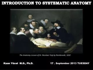
Human Anatomy, First Edition McKinley & O'Loughlin. Chapter 1 Lecture Outline: A First Look at Anatomy . A First Look at Anatomy. Anatomy is the study of structure . The word anatomy is derived from Greek and means “to cut up” or “to cut open.”

E N D
Human Anatomy, First EditionMcKinley & O'Loughlin Chapter 1 Lecture Outline: A First Look at Anatomy
A First Look at Anatomy • Anatomy is the study of structure. • The word anatomy is derived from Greek and means “to cut up” or “to cut open.” • Anatomists examine the relationships among parts of the body along with the structure of individual organs.
Introduction to Anatomy • Physiology • The scientific discipline that studies the function of body structures. • Structure and function cannot be completely separated. • Form is related to function.
Levels of Organization in the Human Body • The simplest level of organization within the body is the chemical level, which is composed of atoms and molecules. • Atoms are the smallest units of matter.
Levels of Organization in the Human Body • Molecules • Two or more atoms combine to form a molecule, such as a protein, a water molecule, or a vitamin. • Macromolecules • Larger and more complex molecules such as DNA and proteins.
Levels of Organization in the Human Body • At the cellular level, specialized structural and functional units called organelles permit all living cells to share some common functions.
Levels of Organization in the Human Body • Large molecules join in specific ways to form cells, the basic units of structure and function in organisms. • The cell is the smallest structural unit that exhibits the characteristics of living things (organisms), and it is the smallest living portion of the human body.
Levels of Organization in the Human Body • Tissues • Groups of similar cells with a common function form tissue. • Tissues are precise organizations of similar cells that perform specialized functions.
Levels of Organization in the Human Body • Organs • Different tissue types that work together to perform specific, complex functions form an organ. • Organ Systems • The organ system level consists of related organs that work together to coordinate activities and achieve a common function. • There are 11 organ systems in the human body.
Levels of Organization in the Human Body • Organism • All body systems function interdependently in a single living human being, the organism.
The Four Types of Tissues in the Human Body Are: • Epithelial tissue covers exposed surfaces and lines body cavities. • Example: The inner lining of the digestive system
The Four Types of Tissues • Connective tissue protects, supports, and interconnects body parts and organs. • Can be solid (such as bone), liquid (such as blood), or intermediate (such as cartilage).
The Four Types of Tissues • Muscle tissue produces movement. • Skeletal muscle • Smooth muscle • Cardiac muscle
The Four Types of Tissues • Nervous tissue conducts impulses for internal communication. • Brain, spinal cord, and nerves
Integumentary • Provides protection • Regulates body temperature • Site of cutaneous receptors • Synthesizes vitamin D • Prevents water loss
Skeletal • Provides support and protection • Site of hematopoeisis (blood cell production) • Stores calcium and phosphorus • Allows for body movement
Muscular • Produces body movement • Generates heat when muscles contract
Nervous • A regulatory system that controls body movement • Responds to sensory stimuli • Helps control all other systems of the body • Also responsible for consciousness, intelligence, memory
Endocrine • Consists of glands and cell clusters that secrete hormones, some of which regulate • body and cellular growth • chemical levels in the body • reproductive functions
Cardiovascular • Consists of a pump (the heart) that moves blood through blood vessels in order to distribute hormones, nutrients, gases, and pick up waste products
Lymphatic • Transports and filters lymph (interstitial fluid) • Initiates an immune response when necessary
Respiratory • Responsible for exchange of gases (oxygen and carbon dioxide) between blood and the air in the lungs
Digestive • Mechanically and chemically digests food materials • Absorbs nutrients • Expels waste products
Urinary • Filters the blood and removes waste products from the blood • Concentrates waste products in the form of urine, and expels urine from the body
Male Reproductive System • Produces male sex cells (sperm) and male hormones (e.g., testosterone) • Transfers sperm to the female
Female Reproductive System • Produces female sex cells (oocytes) and female hormones (e.g., estrogen and progesterone) • Receives sperm from male • Site of fertilization of oocyte • Site of growth and development of embryo and fetus
AnatomicalTerminology • Anatomic position is a specific body position in which an individual stands upright with the feet parallel and flat on the floor. • The head is level, and the eyes look forward toward the observer. • The arms are at either side of the body with the palms facing forward and the thumbs pointing away from the body.
Anatomical Terminology • A plane is an imaginary surface that slices the body into specific sections. • The three major anatomic planes of reference are the coronal, transverse, and sagittal planes.
Sectionsand Planes A coronal plane, also called a frontal plane, is a vertical plane that divides the body into anterior (front) and posterior (back) parts.
Sectionsand Planes • A transverse plane, also called a cross-sectional plane or horizontal plane, cuts perpendicularly along the long axis of the body or organ separating it into both superior (upper) and inferior (lower) parts.
Sectionsand Planes • A sagittal plane or median plane, extends through the body or organ vertically and divides the structure into right and left halves.
Sections and Planes • A sagittal plane in the body midline is a midsagittal plane. • A plane that is parallel to the midsagittal plane, but either to the left or the right of it, is termed a parasagittal (or sagittal) plane. • A minor plane, called the oblique plane, passes through the specimen at an angle.
Directional Terms of the Body • Directional terms are precise and brief, and for most of them there is a correlative term that means just the opposite.
Relative and Directional Terms of the Body • Relative to front (belly side) or back (back side) of the body : • Anterior = In front of; toward the front surface • Posterior = In back of; toward the back surface • Dorsal =At the back side of the human body • Ventral = At the belly side of the human body
Relative and Directional Terms of the Body • Relative to the head or tail of the body: • Superior = Toward the head or above • Inferior = Toward feet not head • Caudal = At the rear or tail end • Cranial = At the head end
Relative and Directional Terms of the Body • Relative to the midline or center of the body: • Medial = Toward the midline of the body • Lateral = Away from the midline of the body • Deep = On the inside, underneath another structure • Superficial = On the outside
Relative and Directional Terms of the Body • Relative to point of attachment of the appendage: • Proximal = Closest to point of attachment to trunk • Distal = Furthest from point of attachment to trunk
Body Regions • The human body is partitioned into two main regions, called the axial and appendicular regions. • the axial region includes the head, neck, and trunk which comprise the main vertical axis of our body • our limbs, or appendages, attach to the body’s axis and make up the appendicular region
Body Cavitiesand Membranes • The posterior aspect of the body has two enclosed cavities • A cranial cavity is formed by the cranium and houses the brain. • A vertebral canal is formed by the individual bones of the vertebral column and contains the spinal cord.
Body Cavities • Both the thoracic and abdominopelvic cavities are lined with thin serous membranes, which are composed of two layers: • A parietal layer lines the internal surface of the body wall. • A visceral layer covers the external surface of organs (viscera) within the cavity. • Between the parietal and visceral layers of the serous membrane is a thin serous cavity, containing a lubricating film of serous fluid.
Body Cavities and Membranes • Constant movement of the organs causes friction. • The serous fluid reduces friction and helps the organs move smoothly against both one another and the body wall.
Body Cavities and Membranes • The median space in the thoracic cavity is called the mediastinum. • It contains the heart, thymus, esophagus, trachea, and major blood vessels that connect to the heart.
Body Cavities and Membranes • Within the mediastinum, the heart is enclosed by a two-layered serous membrane called thepericardium.
The Thoracic Cavity • The right and left sides of the thoracic cavity contain the lungs; they are lined by a two-layered serous membrane called the pleura. • The outer layer is the parietal pleura; it lines the internal surface of the thoracic wall • The inner layer is the visceral pleura; it covers the external surface of the lung • The narrow, moist, potential space between them is called the pleural cavity