Download
1 / 36
370 likes | 700 Vues
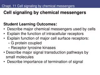
Chapt. 11 Cell signaling by chemical messengers. Cell signaling by chemical messengers Student Learning Outcomes : Describe major chemical messengers used by cells Explain the function of intracellular receptors Explain function of major cell surface receptors: G protein coupled

E N D
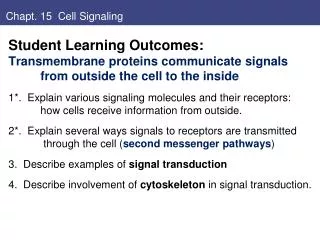
Chapt. 11 Cell signaling by chemical messengers • Cell signaling by chemical messengers • Student Learning Outcomes: • Describe major chemical messengers used by cells • Explain the function of intracellular receptors • Explain function of major cell surface receptors: • G protein coupled • Receptor tyrosine kinases • Describe major signal transduction pathways by small molecules • Describe importance of termination of signal
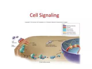
General features of signaling • Chemical messengers include: • hormones, neurotransmitters, cytokines, retinoids, growth factors • Some bind intracellular receptors • Nuclear hormone • Some bind surface receptors • Ion channels, • Tyrosine kinase • G-protein-coupled Second messengers transmit Fig. 11.1
Example of Nicotinic Acetylcholine Receptor • Nicotinic ACh receptor: • High specificity • Nerve signal • Ach vesicles released • Bind ACh receptors on muscle • Opens ion channel • Triggers muscle contraction Fig. 11.2; Acetylcholine receptors at neuromuscular junction
Nicotinic Acetylcholine Receptor • Nicotinic ACh receptor: • 5 subunits, 2 bind ACh • Opens ion channel: • K+ out, Na+ in • Muscle contraciton • Acetylcholinesterase in synaptic cleft stops signal Fig. 11.3
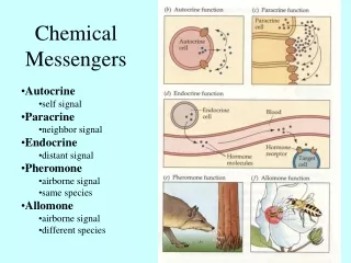
Models of Cell-Cell signaling: Endocrine: Distant targets • Estrogen hormone Paracrine: Local targets • Neurotransmitter Autocrine: self • T cells, cancer cells Fig. 11.4
Some Chemical Messengers • Some chemical messengers: • Nervous system: • Small molecule neurotransmitters • Neuropeptides (4-35 aa) • Endocrine system: • Polypeptide hormones (insulin) • Steroid hormones • Thyroid hormone • Retinoids Fig. 11.5 small molecule neurotransmitters
Some Chemical Messengers • Some chemical messengers: • Immune system: • Cytokines are small proteins • Eicosanoids – prostaglandins • respond to injuries • PGI2 vasodilation • Growth factors – polypeptides • PDGF (Platelet-derived) • EGF (epidermal) Fig. 11.6
II. Intracellular Receptors are Transcription Factors • Properties of messenger determine receptor type: • Hyrophilic hormones bind surface receptors • Lipophilic hormones cross membrane, bind intracellular receptors: • Receptors may be in cytoplasm or nucleus • Often regulate transcription Fig. 11.7
Nuclear hormone superfamily • Steroid hormone-thyroid hormone superfamily: • Nuclear hormone receptors • RAR, TR, VDR, AR • Heterodimers with RXR bind DNA, activate transcription Fig. 11.8
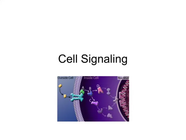
III. Plasma membrane receptors • Plasma membrane receptors function by signal transduction inside cell: • Receptors have membrane-spanning a-helices • Extracellular domain binds messenger • Intracellular domain initiates signal cascade, amplifies signal • Rapid response: ion channels, enzyme activity • Slower effects on gene expression • 1. Ion channel receptors (ACh Figs. 2, 3) • 2. Kinase receptors or bind kinases (RTK) • 3. Heptahelical – G-protein coupled (epinephrine)
Pathways of Intracellular Signal Transduction Intracellular signal transduction: Chain of reactions that transmits signals from cell surface, amplifies, to intracellular targets. Different major mechanisms: cAMP and protein phosphoryation (PKA) cGMP Phospholipids and Ca++ DAG and PKC, IP3 and Ca++, PIP3/AKT Ras, Raf, MAP kinase JAK/STAT; TGFb/Smad
Receptor Tyrosine Kinases and related • 2. Kinases or bind Kinase receptors: signal by first phosphorylating proteins, binding other proteins • *Tyrosine kinase receptors • JAK-STAT receptors bind Januses Kinases • Ser-thr kinase receptors Fig. 11.9
3. Heptahelical Receptors signal by G proteins • 3. Heptahelical receptors signal through heterotrimeric G proteins, second messengers • Binding hormone initiates series of events • GDP-GTP a subunit • Second messengers are small molecules: • cAMP • DAG = diacylglycerol • IP3 = phospatidyl inositol Fig. 11.10
RTK – growth factor receptor and Ras • Tyrosine Kinase Receptor signals through Ras: • Growth factor binds; self-phosphorylation of RTK • Adaptorproteins bind to P-tyr through SH2 domain • Convey signal to membrane-bound Ras • GTP activates Ras (small GTP-binding protein), • Activated Ras binds Raf, signals via MAP kinase pathway Fig. 11.11
Regulation of Ras proteins • Ras-GTP activity is terminated by GTP hydrolysis, stimulated by interaction of Ras-GTP with GTPase-activating proteins. • Ras is mutated in cancers: • Mutated Ras proteins are continuously in active GTP-bound form, driving proliferation of cancer cells in absence of growth factor
Phosphatidyl inositol signaling molecules • Phosphatidylinositol phosphates (PIP) function in signal transduction: • either RTK or heptahelical paths • PI is glycolipid • PI -> PI-4,5-bisP • PLC (phospholipase) -> DAG + IP3 • DAG in membrane activates PKC; • IP3 cytoplasm • PLCg from RTK path • PLCb from G-protein coupled path Fig. 11.12
PLC forms DAG + IP3 • Two forms of phospholipase C: • PLC-β stimulated by G proteins (G-coupled receptors). • PLC-γ has SH2 domains, associates with (RTK). • Tyr phosphorylation increases PLC- γactivity, stimulating hydrolysis of PIP2 to DAG, IP3 • DAG remains in membrane, • Activates protein-ser/thr • kinases of PKC family • (protein kinase C): • Diverse substrates for PKC: • Transcription factors • Actin binding proteins • Phorbol esters activate PKC
IP3 mobilizes Ca2+ • IP3 is small polar molecule released to cytosol; signals release of Ca2+ from ER • IP3 binds receptors that are ligand-gated Ca2+ channels. • Cytosol concentration of Ca2+ maintained at extremely low level by Ca2+ pumps.
RTK Insulin receptor has divergent signaling paths • * Insulin receptor signals through several paths: • Binding of hormone causes autophosphorylation • Binds IRS (insulin receptor substrates), PO4 those: • Grb2 can signal through Ras and MAPK path • Other proteins bind, interact with PIPs in membrane Fig. 11.13 Insulin signaling: PLC - phospholipase PIP – phosphatidylinositol forms
JAK-STAT receptors • JAK-STAT receptors: tyrosine kinase-associated • Often for cytokine signaling – more direct to nucleus • JAK = Janus kinase (just another kinase); • STAT = signal transducer, activator of transcription Fig. 11.15
Receptor ser-thr kinases • Receptor ser-thr kinases for proteins of TGF superfamily • (TGF-b cytokine/ hormone for tissue repair) • Two different membrane-spanning subunits • Smad proteins are receptor-specitic, except Co-smad (Smad4) • Smad complex activates or inhibits transcription Fig. 11.16
Heptahelical receptors use heterotrimeric G proteins • Heptahelical receptors use heterotrimeric G proteins Fig. 11.17
Table 1 Subunits for Heterotrimeric G proteins • Table 11.1 Subunits of Heterotrimeric G-proteins • Many genes (16 a, 5 b, 11 g in mammals) • Ras is also related (small GTP binding protein) • as, Ga(s)Stimulates Adenylyl cyclase • ex. glucagon, epinephrine regulate metabolic enzymes • cholera toxin modifies, keeps it active • ai/o Ga(i/o) Inhibits Adenylyl cyclase • epinephrine, neurotransmitters • pertussis toxin modifies and inactivates • aq11 Ga(q/11) activates PLCb • epinephrine, acetylcholine, histamine
Adenylyl cyclase & cAMP phosphodiesterase • Adenylyl cyclase forms cAMP, second messenger; • cAMP phosphodiesterase cleaves to stop signal Fig. 11.18
cAMP Regulates protein kinase A • Glucagon & epinephrine signal through G-coupled receptorsto increase cAMP • Effects mediated by cAMP-dependent protein kinase, or protein kinase A (PKA) • Inactive form has 2 regulatory, 2 catalytic subunits. • cAMP binds to regulatory subunits, which dissociate. • Free catalytic subunits phosphorylate serine on target proteins Fig. 9.9
PKA stimulates glycogen breakdown: • Ex. PKA stimulates breakdown of glycogen: • PKA phosphorylates 2 enzymes: • Phosphorylase kinase activated, -> activates glycogen phosphorylase. • Glycogen synthase • is inactivated • Glycogen breakdown • stimulated • Glycogen synthesis • blocked.
Cyclic AMP induces gene expression • Increased cAMP can activate transcription of genes • That have regulatory sequence —cAMP response element, or CRE • Free catalytic subunit of PKA goes to nucleus, phosphorylates transcription factor CREB • (CRE-binding protein).
Pathways of Intracellular Signal Transduction cAMP can also directly regulate ion channels: second messenger in sensing smells — odorant receptors are G protein-coupled; stimulate adenylyl cyclase, leading to an increase in cAMP. cAMP opens Na+ channels in plasma membrane, leading to initiation of a nerve impulse.
Phosphatidyl inositol signaling molecules • Phosphatidylinositol phosphates (PIP) function in signal transduction: • either RTK or heptahelical paths • PI -> PI-4,5-bisP • PLC (phospholipase) -> DAG + IP3 • DAG in membrane activates PKC; • IP3 cytoplasm • PLCb from G-protein coupled path Fig. 11.12
IP3 mobilizes Ca2+ • IP3 is small polar molecule released to cytosol; signals release of Ca2+ from ER • IP3 binds receptors that are ligand-gated Ca2+ channels. • Cytosol concentration of Ca2+ maintained at extremely low level by Ca2+ pumps.
Function of Ca2+ and calmodulin • Increased Ca2+ affects activity of several proteins, including protein kinases and phosphatases: • Calmodulin is activated when Ca2+ concentration increases. • Ca2+/calmodulin binds to target proteins, e.g. some protein kinases • CaM kinase family activated by Ca2+/calmodulin; • phosphorylates metabolic enzymes, ion channels, transcription factors, regulate synthesis and release of neurotransmitters.
Termination of signal: • Termination of signal: • Some turn off quickly, others slowly • Many different steps • Diseases from persistence of signal: • Cancer and Ras Fig. 11.19
Key concepts • Cells communicate to integrate cellular functions. • Chemical messages bind receptors on cells (intra-cellular or plasma membrane bound) • Intracellular receptors primarily activate transcription • Plasma membrane receptors are two main types: • Tyrosine kinase and kinase-associated • G-protein-coupled receptors • Various mechanisms for second messenger, can converge from different hormones
Review questions • Pseudohypoparathyroidism is heritable disorder caused by target-organ unresponsiveness to parathyroid hormone (a poplypeptide hormone secreted by the parathyroid gland). One of the mutations that causes this diseases occurs in the gene encoding Gsa in certain cells. • 3. The receptor for parathyroid hormone is most likely which one of the following: • An intracellular transcription factor • B. A cytoplasmicguanylylcyclase • C. A receptor that must be endocytosed in clathrin-coated pits to transmit its signal • D. A heptahelical receptor • E. A tyrosine kinase receptor
review • This mutation (from question 3) likely has which one of the following characteristics? • It is a gain-of-function mutation • It decreases the GTPase activity of the Gas subunit • It decreases synthesis of cAMP in response to parathyroid hormone. • It decreases generation of IP3 in response to parathyroid hormone • It decreases synthesis of phosphatidylinositol 3,4,5-triphosphate in response to parathyroid hormone.
Clinical comments • Mya Sthenia has myasthenia gravis, • autoimmune – Antibodies directed against nicotinic ACh receptor in skeletal muscle. • Fatigue, inability to do repeated tasks; numbers of ACh receptors greatly reduced • Inhibitor of acetylchoinesterase briefly increases muscle strength • Ann O-Rexia - anorexia nervosa. • Endocrine hormones mobilize fuels from adipose tissue • Epinephrine (adrenaline) (GPCR) promotes fuel mobilization • Different receptors on different cells (ex. Glucagon receptors on liver, not on muscle; liver does gluconeogenesis) • Insulin (special RTK) promotes fuel storage