Male Reproductive System Anatomy
630 likes | 706 Vues
Explore the primary and accessory sex organs, hormones, and functions of the male reproductive system. Learn about testes, sperm production, testosterone, and the duct system. Dive deep into the structures like the epididymis, vas deferens, and ejaculatory duct.

Male Reproductive System Anatomy
E N D
Presentation Transcript
Chapter 19Reproductive System Anatomy Male Reproductive System
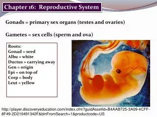
Introduction • Primary Sex Organs • Gonads • Testes – male • Produce sperm – male gamete (sex cell) – exocrine function • Produce testosterone – male hormone – endocrine function • Ovaries – female • Produce Ova/egg – female gamete (sex cell) • Produce estrogen – female hormone • Accessory Sex Organs • Remaining sex organs
Sex Hormones • Testosterone/Estrogen • For the development and functioning of the reproductive organs • For growth and development of other organs and tissues of the body
Male Reproductive OrgansFigure 19.1 Scrotum Fleshy pouch divided into two chambers each housing a testis (male gonads) Extends outside of the body posterior to base of penis Superficial dartos smooth muscle which wrinkles the scrotal surface Deeper skeletal muscle – cremaster Contracts to pull testes closer to the body Sperm need to be cooler than body temperature
Testis Appearance • Olive-size • Covered by capsule – tunica albuginea • Capsule extends in dividing testis into lobules • Lobules contain tightly coiled seminiferous tubules • Sperm producing factories • Empty sperm into rete testis which empty into the epididymis
Interstitial Cells • Surrounding the seminiferous tubules • Produce testosterone – male reproductive hormone • Duct System • Accessory male organs • Transports sperm from the testes through the penis • Epididymis, ductus (vas) deferens, ejaculatory duct, urethra
Male Reproductive Tract • Epididymis • Appearance • Tightly coiled threadlike tube; 20 feet long • Location • On top of the testis, descends along the posterior surface • Epididymis becomes the ductus/vas deferens as it turns up towards the body
Function • Passageway for sperm to the ductus/vas deferens • 2 week journey • Immature sperm and non-motile • Allows time for sperm to mature • Contains cells to reabsorb cellular debris from abnormal or damaged sperm • Contains cells to absorb nutrients from blood • Secretes a substance which prevents premature capicitation • Becoming motile and fully functional • Requires secretion from seminal glands and acidic conditions inside female tract
Ductus (Vas) Deferens • Appearance • Long, winding tube • Location • Continuation of the epididymis • Passes thru the inguinal canal into the abdominal cavity • Arches over the urinary bladder • Enclosed with nerves and blood vessels in connective tissue forming the spermatic cord • Peristaltic contractions empties sperm into the ejaculatory duct which passes through the prostate gland
Function • Store sperm for several months • Transport sperm from to ejaculatory duct • Vasectomy • Small incision into the scrotum cutting through the part of the vas deferens in the scrotum • Sperm are still produced but can no long be expelled out of the body
Ejaculatory DuctFigure 19-5 • Junction of ductus deferens with duct from seminal vesicle • Extends about 1 inch into the prostate gland • Empties sperm into the prostatic urethra
Urethra • Location • Extends from the base of the urinary bladder to the tip of the penis • Last part of the duct system • Regions • Prostatic urethra • Passes through the prostrate gland • Contains the internal urethral sphincter • Membranous urethra • Passes through the muscles of the pelvic floor • Contains external urethral sphincter • Penile urethra • Passes through the length of the penis
Urethral Function • Carries both urine and semen • Semen • Sperm and fluids from the accessory glands • Semen and urine never pass at the same time • During ejaculation, the internal urinary sphincter contracts preventing passage of sperm to bladder and passage of urine to urethra
Accessory Glands • Produce seminal fluid • Seminal vesicles • Prostate gland • Bulbourethral glands • Functions • Contribute fluids of semen – seminal fluids • Nutrients for motility • Activate the sperm • Peristalsis of sperm and fluids • Produce buffers against acidity of urethra and vagina
Seminal Vesicle • Paired tubular glands which attach to the vas deferens at the base of the urinary bladder • Secretes the major portion of the seminal fluid (60%) • Thick, yellowish secretion • Fructose - sugar - energy for the sperm motility • Prostaglandins for peristalsis in male and female tract • Fibrinogen – forming a temporary clot of semen in vagina after ejaculation • Alkaline secretion to neutralize acids • Secretion causes sperm to become motile
Prostate Gland • Appearance • Single gland; chestnut shape • Ejaculatory duct passes through • Location • Surrounds the prostatic urethra • Function • Secretes prostatic fluid – 30% of seminal fluid • Thin, milky, alkaline fluid • Contains an antibiotic that may help prevent urinary tract infections in males • Secretion is released into the urethra
Bulbourethral Glands • Location • Inferior to the prostate gland • Appearance • Very small pea-sized gland • Function • Secretes a clear, thick, sticky, alkaline mucous fluid • Lubricates the penis for sexual intercourse • Cleanse the urethra of traces of acidic urine
Penis Figure 19-6 • Tubular organ, contains penile urethra • Three regions • Root • Attaches penis to body wall • Body/shaft • Contains erectile tissue to deliver the sperm to the female vagina • Spongy tissue that fills with blood causing the penis to enlarge and become rigid – Erection
Glans • Expanded distal end surrounding opening • External urethral meatus • Covered by loose skin • Prepuce (foreskin) • Removed by circumcision
Chapter 19The Reproductive System Anatomy Female Reproductive System
Female Reproductive System • Functions • Produce the female gametes (ova) • Nurture and protect the developing fetus • Produce female sex hormones • Primary reproductive organ – Gonad • Ovaries • Exocrine function – produce eggs/ova • Endocrine function – produce hormones • Estrogens, progesterone
Ovary Figure 19-8 • Appearance • Paired, almond shaped organs • Pale white or yellowish color • Nodular consistency resembling lumpy oatmeal • Location • Suspended by ligaments in the pelvic cavity • Broad ligament; Ovarian ligament • Nourishment/removal of waste • Ovarian artery and vein • Function • Development of egg cells to maturation • About 3 months
Uterine Tubes/Fallopian TubesFigure 19-11 • Location • Extend from the ovaries to the uterus • Appearance • Muscular tube lined with cilia • Expands near ovaries to form funnel shaped structure – infundibulum containing finger-like projections called fimbriae • Does not make physical contact with the ovaries • Fimbriae contain cilia that beat toward the tube forming currents with move the ovum into the tube
Function • Receive the ovulated egg • Depends on movements of the cilia of fimbriae • Some eggs are lost in the peritoneal cavity and might even be fertilized there • If fertilization is to occur, the secondary oocyte must meet the sperm in 12 – 24 hours • Unfertilized oocytes will degenerate • Carry egg (zygote if fertilization occurred) to the uterus • Muscular walls for peristalsis • Rhythmic beating of cilia in uterine tubes
Uterus • Appearance • Hollow, muscular organ • Shape of an inverted pear • Fundus, body, cervix • Cervix - Lower 1/3 of the uterus projecting into the vagina • Location • Between urinary bladder and rectum • Superior to the vagina usually bent forward over the urinary bladder • Held in place in the pelvic cavity by ligaments • Broad ligament
Function • Implantation • Attachment of embryo • Site of embryo development • Prepares each month for zygote • If no fertilization, menstruation occurs
Tissue Layers of Uterus • Endometrium • Inner mucus lining • Two layers • Superficial functional layer • Undergoes changes due to sex hormone levels • Deeper basilar layer • Reponsible for reforming the functional layer monthly • Embryo burrows into this lining – implantation • Sloughs off about every 28 days if fertilization does not occur
Myometrium • Thick muscular layer • Contracts during childbirth • Perimetrium • Outer layer • Visceral peritoneum
Vagina • Appearance • Elastic, muscular tube; 3-4 inches long • Opening is the vaginal orifice covered by the hymen • Contains resident bacterial supported by nutrients in mucus of vagina • pH is 3.5 – 4.5 restricts growth of pathogens • Location • Extends from the uterus to the outside • Posterior to the bladder/Anterior to the rectum
Functions • Transports uterine secretions • Transports the fetus during childbirth – birth canal • Receives the penis during intercourse
External Genitalia Figure 19-12 • Female reproductive structures external to the vagina • Also called the Vulva • Mon pubis, labia, clitoris, vestibular glands • Mons pubis • Fatty rounded area over the pubic symphysis • Covered with pubic hair after puberty
Labia • Labia majora • Hair covered skin folds • Labia minora • Located between the labia majora • Hairless • Clitoris • Small projection at anterior end of vulva • Corresponding to penis of the male • Hooded by the prepuce • Contains erectile tissue which becomes swollen with blood during sexual excitement
Vestibular glands • Produce mucus • Lubricates distal end of vagina during intercourse
Mammary Glands Figure 19-13 • Glands of the breast secreting milk in a process called lactation • Breast is divided into lobes each containing ducts which converge to a single lactiferous duct • Near the nipple, the lactiferous ducts expands into the lactiferous sinus • Open onto surface of the nipple • Nipple surrounded by reddish-brown tissue called areola containing sebaceous glands • Breast is connected to chest muscle wall by suspensory ligaments
Chapter 19Reproductive System Physiology of the Reproductive System
Human Life Cycle Figure 16.4 • Somatic body cells contain 46 chromosomes – diploid number or 2n • Sex cells – gametes – haploid number or n • Sperm cell – 23 chromosomes • Egg cell – 23 chromosomes • Fertilization • Union of sperm and egg produces a zygote with 46 chromosomes • Half the characteristics from male sex cell • Half the characteristics from female sex cell
Meiosis • Special type of division which occurs in gonads – ovaries, testes • Production of gametes with n number of chromosomes • Reduces the diploid number of chromosomes to the haploid number • Oogenesis • Production of the female sex cells – ova • Spermatogenesis • Production of the male sex cells - spermatozoa
Meiosis • Consists of two successive divisions of the nucleus • Meiosis I and Meiosis II • Each division is divided into stages • Prophase, metaphase, anaphase, telophase • Results in 4 daughter cells (gametes) instead of 2 • A way to reduce the number of chromosomes in half
Spermatogenesis Figure 19-3 • Sperm production • 3 processes • Mitosis • Stem cells – spermatogonia • Beings during puberty and continues throughout life • Millions of sperm produced daily • Occurs in the seminiferous tubules • One daughter cell remains in seminiferous tubule; the other is pushed into the lumen • Primary Spermatocyte
Meiosis I • Meiosis I • Chromosomes replicate • Homologous pairs – maternal and paternal come together in a process called synapsis • 4 chromosomes called a tetrad • Crossing over of genetic information may occur • At the end of meiosis I daughter cells receive both copies of either the maternal chromosome or the paternal chromosome from each tetrad • Forms the secondary spermatocytes
Meiosis II • Each secondary spermatocyte contains 23 chromosomes but each consists of 2 chromatids • Duplicate chromatids will separate in meiosis II • Forms the spermatids • Each with 23 single chromosomes • n number of chromosomes
Spermiogenesis Figure 16.5b • Process of the last stage of sperm development • Excess cytoplasm is sloughed off • Sperm is compacted into three regions – head, midpiece and tail • Tail – flagella develops
Mature Sperm Figure 19-4 • Head • Nucleus containing DNA – 23 chromosomes • Covered with an acrosome • Similar to a large lysosome • In close contact with the oocyte the acrosomal membrane breaks down and releases enzymes that help the sperm penetrate through the follicle cells surrounding the oocyte
Midpiece • Contains centrioles which contain filaments that form the flagella • Filaments are covered by mitochondria providing he energy for movements of the flagella • Tail • Flagella • Only example of a flagellum in humans • Enable sperm to move long distances in a short time
Hormones • FSH stimulates the seminiferous tubules to produce sperm • LH luteinizing hormone/ISCH interstitial cell stimulating hormone • Testosterone Production • Function of the interstitial cells in seminiferous tubules • Testosterone • Stimulates reproductive organs to develop • Functions in the sex drive • Causes the secondary male sex characteristics to appear
Male Secondary Sex Characteristics • Deepening of voice due to enlargement of larynx • Increased hair growth all over the body • Axillary regions • Pubic regions • Face • Enlargement of skeletal muscles • Thickening of bones
Oogenesis Figure 19-9 • Process of production of female gametes – ova • Total number of eggs a female can release is determined by the time she is born • Release of eggs begins during puberty and ends in her 50’s or earlier • Menopause – gradual decline and end to a woman’s ability to reproduce
Oogonia – female stem cells • Somatic cells containing the 2n number of chromosomes • These cells are located in the periphery of the ovary • These cells go through mitosis in the female FETUS • Daughter cells are called primary oocytes – in prophase I of meiosis • Primary ooctye is pushed into the ovary connective tissue and is surrounded by follicle cells
At birth all the cells are primary oocytes • Oogonia no longer exist • This the females’ life supply of eggs approximately 2 million • Waiting to undergo meiosis and produce functional eggs • Remain at this point until puberty
Meiosis • Production of 4 daughter cells but the cytoplasm is not evenly distributed • One functional ovum with cytoplasm • 3 nonfunctional polar bodies which disintegrate • Ovary releases a secondary oocyte not a mature ovum • Meiosis II does not occur producing a mature ovum unless fertilization occurs