Pleural Disease
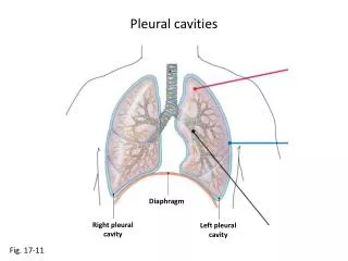
Pleural Disease. Pleural Disease. Air in the pleural cavity Pneumothorax: from airway or from atmosphere. Pneumothorax. Air in the pleural space Expiratory view and lateral decubitus view (affected side up) are best for evaluation


Pleural Disease
E N D
Presentation Transcript
Pleural Disease • Air in the pleural cavity • Pneumothorax: from airway or from atmosphere
Pneumothorax • Air in the pleural space • Expiratory view and lateral decubitus view (affected side up) are best for evaluation • Commonly due to rupture of blebs or bullae (emphysema) of the lung or direct trauma (rib fractures, knife or bullet wounds)
Pneumothorax • X-ray signs: • Lucent area with no lung markings located laterally or at the apex of thoracic cavity (if the amount of air is small) • The border of collapsed lung is seen as a sharp line (visceral pleura)
Bilateral Pneumothoraces • PA film inspiration • Lucent area adjacent to lateral chest walls (arrows) • No lung vessels seen (beyond visceral pleura) • Pleural border of collapsed lungs vaguely seen
Bilateral Pneumothoraces • Expiration PA film • Pneumothoraces appear larger because lung volume is decreased while pleural volume is unchanged
Massive Left Pneumothorax • PA chest • Very large left pneumothorax • Left chest lucent • Collapsed left lung, opaque area at the left hilum • Mediastinum normal (no shift)
Tension Pneumothorax • X-ray signs: • In tension pneumothorax a ball-valve mechanism will cause increased air pressure which will displace the mediastinum away from the side of the tension pneumothorax and flatten or invert the hemidiaphragm on the side of the pneumothorax • Surgical emergency
Tension Pneumothorax • Lucent periphery • Pleural margin • Inverted diaphragm • Shift of midline to the left • Atelectasis / loss of volume on left