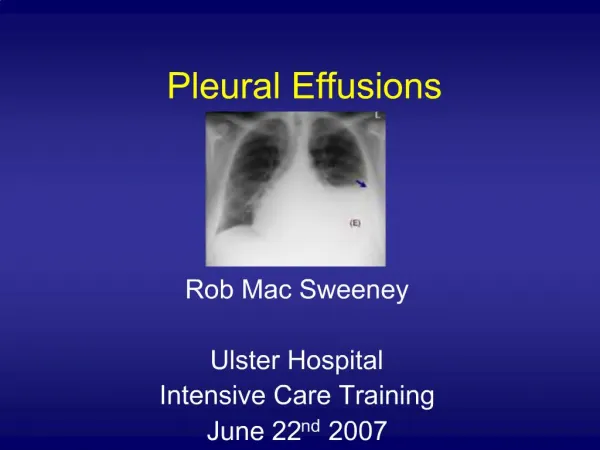
Pleural Effusions
Learn about the physiology of the normal lung, pleural effusion pathophysiology, causes, symptoms, diagnosis, and treatment options. Understand the layers of the lung and pleura fluid dynamics.

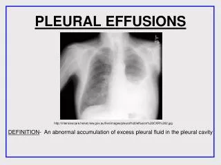
Pleural Effusions
E N D
Presentation Transcript
Pleural Effusions Kady Rejret, RN,BSN Alverno College MSN-621
Navigating this tutorial Takes you back to the Outcomes page Takes you to next page Takes you to previous page Takes you to previous page viewed
OUTCOMESClick on the topic below you would like to view • Describe the pathophysiology of the normal lung • Describe the pathophysiology of a pleural effusion • Describe the main causes of a pleural effusion • Differentiate among the manifestations of fluid collections • Describe the signs and symptoms of a pleural effusion • Explain diagnostic methods • Describe the various treatment options
Normal lung pleural effusion Picture used with permission (Allibone, 2006, p.56)
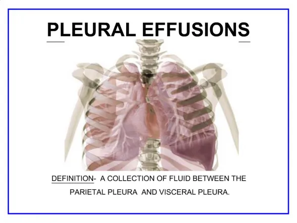
Physiology of the normal lung • The lungs are soft, spongy, cone-shaped organs located in the chest cavity. • They are separated by the mediastinum and the heart. • There are 3 lobes on the right lung and 2 lobes on the left lung.
Pleura -serous fluid that allows for the parietal pleura (outer lining) and visceral pleura (inner lining) to glide over each other without separation(Porth, 2005, p. 639) -contains about 5-15ml of fluid at one time -Pleural fluid is produced by the parietal pleura and absorbed by the visceral pleura as a continuous process.(Drummond Hayes, 2001, p. 32) -about 100-200ml of fluid circulates though the pleural space within a 24-hour period(Brubacher & Holmes Gobel, 2003) -has an alkaline pH of about 7.64(Drummond Hayes, 2001, p. 33)
Layers of the lung Pleural Space • thin, transparent, serous membrane which lines the thoracic cavity • a potential space between the parietal pleura and visceral pleura Rib Cage Lung Picture used with permission Allibone, 2006
Layers of the lung Parietal Pleura • Lines the thoracic cavity, including the thoracic cage, mediastinum, and diaphragm • Contains sensory nerve endings that can detect pain Rib Cage Lung Picture used with permission Allibone, 2006
Layers of the lung Visceral Pleura • Lines the entire surface of the lung • Contains NO sensory nerve endings that detect pain Rib Cage Lung Picture used with permission Allibone, 2006
Review question: Pleuritic chest pain indicates inflammation or irritation of the parietal pleura or visceral pleura? (click on the correct answer)
Think again! The visceral pleura contains no nerve endings for detecting pain.
Correct! The parietal pleura contains sensory nerve endings that can detect pain.
Review question: The pleural space typically contains how much fluid? 5-15ml 50-100ml 100-200ml
Think again! about 100-200ml of fluid circulates though the pleural space within a 24-hour period
Correct! • 5-15ml of fluid are present at one time • The pleural space is a potential space between the parietal pleura and visceral pleura, allowing them to glide over each other without separation
The lungs are supplied with blood via the pulmonary and bronchial circulations. Pulmonary circulation: supplied from the pulmonary artery and provides for gas exchange function of the lungs. Bronchial circulation: distributes blood to the conducting airways and supporting structures of the lung. The normal lung
Intrapulmonary pressure -the pressure within the alveoli -as the chest expands on inspiration the intrapulmonary pressure becomes more negative, which causes air to be sucked into the lungs. (Allibone, 2006, p. 56) Intrapleural pressure -Negative pressure is created in the pleural space as the thoracic cage enlarges and the lungs recoil during normal inspiration -negative pressure may be lost if fluid collects in the pleural space, making the lung unable to expand fully. (Allibone, 2006, p. 56) The normal lung
The normal lung • cells within the pleura are primarily mesothelial cells that line the surfaces of the pleural membranes and some white blood cells (WBC). • The visceral pleura absorbs fluid, which then drains into the lymphatic system and returns to the blood • Protein in the circulation and balanced pressures keep excessive amounts of fluid from seeping out of the blood vessels into the pleural space (Pumonary Channel, 2007)
Let’s review Click on the words below to send them to their correct position within the diagram. Rib cage Lung Pleural Space Visceral Pleura Parietal Pleura Picture used with permission Allibone, 2006
Let’s review Fluid is absorbed by the: Parietal Pleura Pleural Space Visceral Pleura
Think Again - - - • Pleural fluid is produced by the parietal pleura • The pleural space is a potential space between the parietal pleura and visceral pleura • Negative pressure is created in the pleural space
C o r r e c t ! ! ! • Pleural fluid is produced by the parietal pleura and absorbed by the visceral pleura as a continuous process. • The visceral pleura absorbs fluid, which then drains into the lymphatic system and returns to the blood
OUTCOMESClick on the topic below you would like to view • Describe the pathophysiology of the normal lung • Describe the pathophysiology of a pleural effusion • Describe the main causes of a pleural effusion • Differentiate among the manifestations of fluid collections • Describe the signs and symptoms of a pleural effusion • Explain diagnostic methods • Describe the various treatment options
Pleural effusion • Created by an abnormal collection of fluid in the pleural space • Seen in chest X-ray with presence of about 200ml pleural fluid • Fluid in X-ray seen as a dense, white shadow with a concave upper edge (fluid level) (Allibone, 2006) Click on the pleural effusion in the picture! Used with permission (Allibone, 2006, p. 59)
Pleural Effusion Fluid accumulates in the pleural space by three mechanisms: -increased drainage of fluid into the space -increased production of fluid by cells in the space -decreased drainage of fluid from the space (pulmonary channel, 2007)
Pleural Effusion • The build-up of fluid presses on the lung, making it difficult for the lung to expand fully. • Part or all of the lung may then collapse (National Cancer Institute, 2007)
Pleural Effusion • Your lungs contain millions of small, elastic air sacs called alveoli • Normally, with each breath the air sacs take in oxygen and release carbon dioxide • Sometimes increased pressure in the blood vessels in your lungs forces fluid into the air sacs, filling them with fluid and preventing absorption of oxygen. (Mayo Foundation for Medical Education and Research, 2006)
Pleural Effusions Malignancy accounts for about 40% of symptomatic pleural effusions, with congestive heart failure and infection being the other leading causes (National Cancer Institute, 2006)
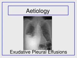
Fluid collection in both lower lobes of the lungs due to CHF Picture used with permission (Allibone, 2006, p. 59)
Main causes of a Pleural Effusion • Congestive Heart Failure (CHF) • Liver failure • Infection • Atelectasis • Cancer • Trauma Click on home icon when finished viewing these topics
Congestive Heart FailureCHF • As the heart fails, pressure in the vein going through the lungs starts to rise. • Due to the heart’s inability to move blood from the pulmonary circulation into the arterial side of systemic circulation, there is a decrease in cardiac output, an increase in left atrial and ventricular end-diastolic pressures, and congestion in the pulmonary circulation. • As the pressure increases, fluid is pushed into the air spaces (alveoli) • This fluid then leaks from the alveoli into the pleural space • This fluid creates a pleural effusion and interrupts normal oxygen movement through the lungs, resulting in shortness of breath
CHF • CHF is the most common cause of pleural effusion. • Frequently the effusions are bilateral (approximately 75% of the time) but may occur alone on either side with the right side being more common. • Fluid is usually straw colored, with low white blood cell counts (<500 cells/mm3) and a mononuclear cell predominance. • With severe congestive heart failure, fluid may persist in spite of vigorous diuresis. (National Lung Health Education Program, 2000) Back
Liver Failure • Negative intrapleural pressure may lead to a transudative effusion due to peritoneal fluid from ascites moving across the diaphragm into the chest (Current Therapy, 2001, p. 208)
Infection • Pneumonia -inflammation of the lung structures, specifically the alveoli and bronchioles • WBCs accumulate in response to infection and inflammation leading to empyema
Atelectasis • Atelectasis is an incomplete expansion of the lung which leads to collapse of the alveoli • Increased negative intrapleural pressure can lead to the collection of fluid in the portion of the lung which is not expanding • This can cause an effusion by fluid leaking out of the lung and into the chest cavity • Atelectasis typically leads to small pleural effusions not requiring surgical intervention
Cancer • Impaired lymphatic drainage of the pleural space due to obstruction by a tumor • Typically due to the interference with the visceral pleura (which absorbs pleural fluid) • A tumor can obstruct pulmonary veins, preventing fluid from being reabsorbed into the bloodstream • A tumor can perforate the thoracic duct • Shedding of malignant cells into the pleural space, decreasing reabsorption of pleural fluid back into the lymphatic system (Brubacher & Holmes Gobel, 2003, p. 1)
Trauma • Increased capillary permeability as a result of inflammation • Fluid (most often, blood) may collect in the lung cavity as a result of trauma to the lung
Pleural fluid types • Transudate • Exudate • Empyema • Chyle • Hemothorax Click on home icon when finished viewing these topics
Transudate • Clear, pale yellow, watery substance • Influenced by systemic factors that alter the formation or absorption of fluid • Increase in hydrostatic pressure • Decrease in plasma oncotic pressure • Contains few protein cells • Common causes: CHF and liver or kidney disease
Exudate • Pale yellow and cloudy substance • Influenced by local factors where fluid absorption is altered (inflammation, infection, cancer) • Rich in protein (serum protein greater than 0.5) • Ratio of pleural fluid LDH and serum LDH is >0.6 • Pleural fluid LDH is more the two-thirds normal upper limit for serum • Rich in white blood cells and immune cells • Always has a low pH • Common causes: pneumonia, cancer, and trauma
Empyema • Pus • Yellow, cloudy, and foul odor • Most likely due to pneumonia, lung abscess, infected chest wounds • Has a pH > 7.2 (Drummond Hayes, 2001, p. 33)
Chyle • Milky fluid • Consists of lymph and fat • Chyle leaks from the thoracic duct -due to lymphatic obstruction (tumor) or trauma • High triglyceride levels found in fluid analysis
Hemothorax • Blood • Usually results from chest injury • A blood vessel ruptures into the pleural space or a bulging area into the aorta (aortic aneurysm) leaks blood into the pleural space • Can occur as a result of bleeding from the ribs, chest wall, pleura, and the lung
Let’s review • Which is NOT a type of fluid that may cause a pleural effusion? -empyema -chylothorax -pneumothorax -hemothorax
This is a fluid that may cause a Pleural Effusion • Empyema (pus), Chylothorax (chyle), and hemothorax (blood) are all fluids that may result in a pleural effusion.
Correct, this is not a fluid! • Pneumothorax is a collection of air in the pleural cavity.
Signs and symptoms • Dyspnea • Cough, usually non-productive • Pleuritic chest pain • Chest pressure • Hypoxemia • Decreased breath sounds on the affected side • Some people may exhibit no symptoms!
Diagnosis • Chest radiograph (x-ray) -able to distinguish >200ml of fluid • Chest ultrasound -locates small amounts or isolated loculated pockets of fluid -able to give precise position of accumulation • Computed Tomography (CT) scan -Differentiates between fluid collection, lung abcess, or tumor
Diagnosis Fluid analysis confirms a pleural effusion Normal pleural fluid has the following characteristics: • clear ultrafiltrate of plasma • pH 7.60-7.64 • protein content less than 2% (1-2 g/dL) • fewer than 1000 WBCs per cubic millimeter • glucose content similar to that of plasma • lactate dehydrogenase (LDH) level less than 50% of plasma and sodium • potassium and calcium concentration similar to that of the interstitial fluid (Abrahamian, 2005, p. 2 of 28)
Non-surgical Treatment Options • Thoracentesis • tPA • Chemical Pleurodesis • Pleurx catheter