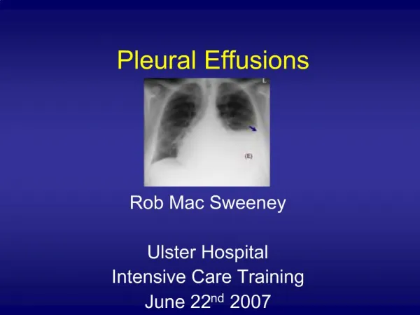
Pleural Effusions
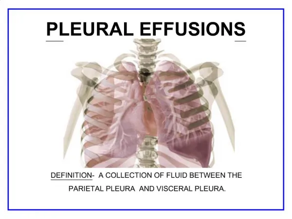
Pleural Effusions. Kara Lee Gallagher USC School of Medicine. Definition. Increased amount of fluid within the pleural cavity Stedman’s Medical Dictionary Accumulation of fluid between the layers of the membrane that lines the lungs and the chest cavity Medline Plus. Epidemiology.

Pleural Effusions
E N D
Presentation Transcript
Pleural Effusions Kara Lee Gallagher USC School of Medicine
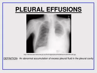
Definition • Increased amount of fluid within the pleural cavity • Stedman’s Medical Dictionary • Accumulation of fluid between the layers of the membrane that lines the lungs and the chest cavity • Medline Plus
Epidemiology • United States • 1 million cases annually • Internationally • 320/100,000 in industrialized countries
Pathophysiology • Normal: 1 mL of pleural fluid • Balance between hydrostatic/oncotic forces and lymphatic drainage • Abnormal: Pleural effusion • Disruption of balance
Clinical History • Dyspnea • Chest pain
Physical Exam • Decreased breath sounds • Dullness to percussion • Decreased tactile fremitus • Egophony • Pleural friction rub
Types • Hydrothorax • Hemothorax • Chylothorax • Pyothorax or Empyema
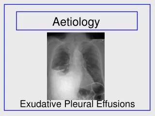
Classification • Transudate • Ultrafiltrate of plasma • Small group of etiologies • Exudate • Produced by host of inflammatory conditions • Large group of etiologies
Workup: Thoracentesis • Light’s criteria: Transudate vs. Exudate • Pleural fluid protein / serum protein > 0.5 • Pleural fluid LDH / serum LDH > 0.6 • Pleural fluid LDH > 2/3 ULN serum LDH
Workup: Thoracentesis • Other criteria: Transudate vs. Exudate • Pleural fluid LDH > 0.45 ULN serum LDH • Pleural fluid cholesterol > 45 mg/dL • Pleural fluid protein > 2.9 g/dL
Workup: Laboratory • LDH > 1000 IU/L • Empyema, Malignancy, Rheumatoid • Glucose < 30 mg/dL • Empyema, Rheumatoid • Glucose between 30 – 50 mg/dL • Lupus, Malignancy, TB
Workup: Laboratory • Lymphocytes > 85% • Chylothorax, Lymphoma, Rheumatoid, TB • Lymphocytes between 50 – 70% • Malignancy • Mesothelial cells > 5% • TB unlikely • ADA > 43 U/mL • Supports TB
Workup: Imaging • Upright Chest X-Ray • Blunting of costophrenic angles • Supine Chest X-Ray • Increased density over lower lung fields • Lateral decubitus Chest X-Ray • Layering
Workup: Imaging • Ultrasound • Aids in identification of loculated effusions • Aids in differentiation of fluid from fibrosis • Aids in identification of thoracentesis site • Available at bedside
Workup: Imaging • CT Scan • Aids in differentiation of • Lung consolidation vs. Pleural effusion • Cystic vs. Solid lesions • Peripheral lung abscess vs. Loculated emypema • Aids in identification of • Necrotic areas • Pleural thickening, nodules, masses • Extent of tumor
Treatment • Treat underlying etiology • Therapeutic thoracentesis
Questions? Image sources cited in notes