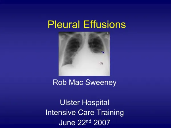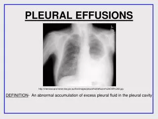
Pleural Effusions
Pleural Effusions. The Pleura. Pleural Pathophysiology. Transpleural pressure imbalance Increased capillary permeability Impaired lymphatic drainage Transdiaphragmatic movement of fluid Pleural effusions of extravascular origin (chylothorax). On CXR. Differential Diagnosis.


Pleural Effusions
E N D
Presentation Transcript
Pleural Pathophysiology • Transpleural pressure imbalance • Increased capillary permeability • Impaired lymphatic drainage • Transdiaphragmatic movement of fluid • Pleural effusions of extravascular origin (chylothorax)
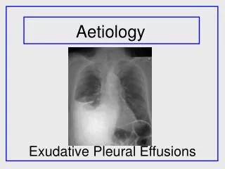
Differential Diagnosis • Transpleural pressure imbalance • CHF • Increased capillary permeability • PNA • Impaired lymphatic drainage • Malignancy • Late PNA (fibrin) • Transdiaphragmatic movement of fluid • Hepatic Hydrothorax • Peritoneal dialysis • Pleural effusions of extravascular origin (chylothorax)
Differential Diagnosis - Complete CHF Hepatic Hydrothorax PD Pancreatitis Lung/Liver abscess Chylous ascites Malignancy Meig’s syndrome (ascites, benign ovarian tumor) Parapneumonic Pulmonary embolism TB Hypoalbuminemia/Nephrotic syndrome Atelectasis/Trapped Lung Asbestosis Rheumatoid lung Yellow Nail Syndrome Duropleural fistula SVC obstruction Sarcoidosis Esophageal perforation Lupus pleuritis Constrictive pericarditis Post-cardiac surgery syndrome
Rule of Thumb – Treat underlying disease CHF Hepatic Hydrothorax PD Pancreatitis Lung/Liver abscess Chylous ascites Malignancy Meig’s syndrome (ascites, benign ovarian tumor) Parapneumonic Pulmonary embolism TB Hypoalbuminemia/Nephrotic syndrome Atelectasis/Trapped Lung Asbestosis Rheumatoid lung Yellow Nail Syndrome Duropleural fistula SVC obstruction Sarcoidosis Esophageal perforation Lupus pleuritis Constrictive pericarditis Post-cardiac surgery syndrome
Thoracentesis – Diagnostic and Therapeutic From UpToDate… INDICATIONS — Pleural effusions are usually detected by physical examination and then confirmed radiographically. Most patients who have a pleural effusion should undergo diagnostic thoracentesis to determine the nature of the effusion (ie, transudate, exudate) and to identify potential causes (eg, malignancy, infection).
Thoracentesis – Diagnostic and Therapeutic What to send?
Thoracentesis – Diagnostic and Therapeutic • What to send? • Cell count • Cytology • pH/glucose • Amylase • Triglycerides • ADA • Gram and Culture (bacterial, viral, fungal, AFB)
Thoracentesis – Diagnostic and Therapeutic Reexpansion Pulmonary Edema If more than 1 liter of pleural fluid is removed at a time during a thoracentesis or from a chest tube RPE may result. RPE may present as asymptomatic radiographic changes or as complete cardiopulmonary collapse. Mortality rate is 20%.
Pleural Fluid Diagnostics • Transpleural pressure imbalance (transudate) • Increased capillary permeability (exudate) • Impaired lymphatic drainage (exudate) • Transdiaphragmatic movement of fluid (transudate) • Pleural effusions of extravascular origin (either)
Pleural Fluid Diagnostics • Transpleural pressure imbalance (transudate) • Increased capillary permeability (exudate) • Impaired lymphatic drainage (exudate) • Transdiaphragmatic movement of fluid (transudate) • Pleural effusions of extravascular origin (either) • Transudates are caused by: • Increased Starling forces • Increased systemic capillary forces (increased rate of filtration) • Increased systemic venous HTN (not really) • Pulm venous HTN (CHF) • Fistula or increased compartment pressure • Exudates are caused by: • Impaired protein and cell clearance from pleural space • Leaky mesothelium
Pleural Fluid Diagnostics • Light’s Criteria: Effusion is likely exudative if at least one of the following exists: • The ratio of pleural fluid protein to serum protein is greater than 0.5 • The ratio of pleural fluid LDH and serum LDH is greater than 0.6 • Pleural fluid LDH is greater than 0.7 times the normal upper limit for serum • Lights diagnosis approx 20% of transudates as exudates. • Modified Light’s Criteria: Effusion is likely exudative if at least one of the following exists: • The ratio of pleural fluid protein to serum protein is greater than 0.5 • The pleural fluid LDH is greater than 0.67 the upper limit of normal serum concentration
Pleural Fluid Diagnostics Exudate Characteristics Usually > 1000 nucleated cells >50,000 nucleated cells is indicative of empyema < 5000 nucleated cells with mononuclear predominance indicated TB >80% lymphocytes indicative of transplant rejection, lymphoma, post-CABG, sarcoid, TB, fungal infection, yellow nail syndrome >10% eosinophils indicative of abestosis, carcinoma, churg-strauss, hemothorax, lymphoma, parasites, PE, sarcoid, TB