Download
1 / 1
20 likes | 195 Vues
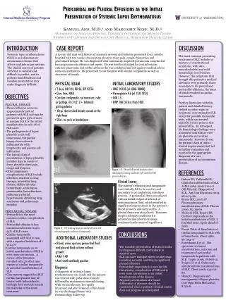
Samuel Ash, M.D. 1 and Margaret Neff, M.D. 2 1 Department of Internal Medicine, University of Washington Medical Center 2 Division of Pulmonary and Critical Care Medicine, Harborview Medical Center. INTRODUCTION. CASE REPORT. DISCUSSION.

E N D
Samuel Ash, M.D.1 and Margaret Neff, M.D.21Department of Internal Medicine, University of Washington Medical Center 2Division of Pulmonary and Critical Care Medicine, Harborview Medical Center INTRODUCTION CASE REPORT DISCUSSION Systemic lupus erythematosus (SLE) is an inflammatory autoimmune disease that affects multiple organ systems. The clinical course of patients with SLE is variable and difficult to predict, and its protean manifestations and variable presentation may make diagnosis difficult. A 19 year old man with history of anorexia nervosa and bulemia presented to an outside hospital with two weeks of worsening pleuritic chest pain, cough, rhinorrhea and generalized fatigue. He was diagnosed with community acquired pneumonia complicated by parapneumonic effusion and sepsis. He was briefly intubated for central venous catheter placement, but within 48 hours he was extubated and left against medical advice with oral antibiotics. He presented to our hospital with similar complaints as well as shortness of breath. The most common presenting syndrome of SLE includes a mixture of constitutional symptoms, with skin, musculoskeletal and mild hematologic involvement. However, the symptoms that brought this patient to medical attention were primarily those secondary to his pleural and pericardial effusions, the latter of which resulted in cardiac tamponade. Further discussion with this patient and detailed history yielded no other signs or symptoms concerning for SLE except for possible monocular iritis, which was treated topically 3 years prior to this presentation. In retrospect, his hematologic findings were consistent with SLE as were his pleuritis and cardiac tamponade. However it was the patient’s lack of initial clinical improvement that led to further evaluation and resulted in the appropriate diagnosis of a rare presentation of an uncommon disease. CONCLUSIONS • The variable presentation of SLE can make its diagnosis difficult, particularly in atypical cases • SLE can have multiple effects on the lungs including serositis resulting in significant pleural effusions • Pericardial tamponade is a rare but life threatening complication of SLE and is even more uncommon as an initial manifestation of the disease • Atypical presentations of a broad differential of diseases should be considered when a patient’s clinical course does not progress as anticipated Pericaridaland Pleural Effusions as the Initial Presentation of Systemic Lupus Erythematosus INITIAL LABORATORY STUDIES ADDITIONAL LABORATORY STUDIES PHYSICAL EXAM • WBC 10120 (nl 4300-10000) • Hemoglobin 9.6 (nl 13.0-18.0) • INR 1.5 • BNP 186 (nl less than 100) • Blood, urine, sputum, pericardial fluid and pleural fluid cultures without growth • ANA 1:40 • Anti-smith antibody positive • T 36.6, HR 124, RR 26, BP 92/56 • Gen: thin, NAD • Cardiac: tachycardic. no murmurs, rubs or gallops. nl s1/s2. 2+ bilateral pitting edema • Resp: diminished breath sounds at the right base • Skin: no rash or breakdown OBJECTIVES • PLEURAL DISEASE • Pleural effusion occurs in approximately 30% of patients with SLE and may be present in up to 93% of cases at autopsy but it is the initial manifestation in only 1% of SLE cases • The pathogenesis of lupus pleuritis is not well understood, but pleural biopsy shows chronic inflammation with lymphocyte and plasma cell infiltration • The typical clinical presentation of lupus pleuritis includes days to weeks of fever, pleuritic chest pain, cough and dyspnea • Other pulmonary complications of SLE include infection, reaction to drug therapy, interstitial lung disease, diffuse alveolar hemorrhage, acute lupus pneumonitis, bronchiectasis, pulmonary arterial hypertension, shrinking lung syndrome and pulmonary embolus • PERICARDIAL DISEASE • Pericarditis is the most common cardiac complication of SLE • Pericardial effusion is less common and occurs in 9 to 54% of SLE cases • Cardiac tamponade associated with SLE is rare, with a reported incidence of 1 to 2.5% • Cardiac tamponade as an initial manifestation of SLE is even more uncommon. A review of the literature revealed only 26 other case reports of cardiac tamponade as an initial manifestation of SLE • Case reports suggest that SLE cardiac tamponade may be treated with indomethacin, but high dose steroids remain the mainstay of the acute treatment Figure 1-2: PA and left lateral decubitus chest radiographs showing moderate right and small left pleural effusions REFERENCES • Dubois EL, Tuffanelli DL. Clinical manifestations of SLE. JAMA 1964; 190(2):104-11. • Gill JM et al. Diagnosis of SLE. Am Fam Physician 2003; 68:2179-86. • Keane MP, Lynch JP. Pleuropulmonary manifestations of SLE. Thorax 2000; 55:159-66. • Mohseni MM, Rogers ER. Cardiac tamponade as the initial manifestation of SLE. J Em Med 2010. Epub ahead of print. • Porcel JM et al. Resolution of cardiac tamponade in SLE with indomethacin. Chest 1989; 96:1193-4. • Rosenbaum E et al. The spectrum of clinical manifestations, outcome and treatment of pericardial tamponade in patients with SLE. Lupus 2009; 18:608-12. • Swigris JJ et al. Pulmonary and thrombotic manifestations of SLE. Chest 2008; 133:271-80. • Wang D. Diagnosis and management of lupus pleuritis. Curr Opin Pulm Med 2002; 8:312-6. Clinical Course Diagnosis A diagnosis of systemic lupus erythematosus was made and the patient was treated with pulse dose steroids followed by maintenance steroid dosing. With steroid therapy, he rapidly improved and after removal of his drains he was discharged home with rheumatology follow-up. The patient’s effusions and tamponade were initially felt to be reactive and secondary to an underlying infectious process. A pericardial drain was placed with an initial output of 480 mL of serosanguinous fluid,which resulted in significant improvement in the patient’s blood pressure and tachycardia. A pleural drain was also placed. However, despite adequate antibiosis his pericardial and pleural drainage continued and remained significant. Figure 3: TTE showing large pericardial effusion with echocardiographic evidence of tamponade.