
The Respiratory System
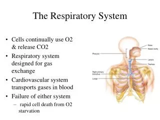
The Respiratory System. Cells continually use O2 & release CO2 Respiratory system designed for gas exchange Cardiovascular system transports gases in blood Failure of either system rapid cell death from O2 starvation. two nasal cavities with bony outgrowths = nasal conchae (superior,

The Respiratory System
E N D
Presentation Transcript
The Respiratory System • Cells continually use O2 & release CO2 • Respiratory system designed for gas exchange • Cardiovascular system transports gases in blood • Failure of either system • rapid cell death from O2 starvation
two nasal cavities with bony outgrowths = nasal conchae (superior, • middle, inferior) • nasal cavities separated by nasal septum and nasal bone • lined with: pseudostratified ciliated epithelial cells - mucus production • also are receptors for odors -lead to nerves -> brain (smell) • lacrimal glands drain into nasal cavities • nasal cavities communicate with cranial sinuses (air-filled chambers within the skull) • nasal cavities empty into the nasopharynx - upper portion of the • pharynx • functions: warm, moisten, and filter incoming air Nose -- Internal Structures • Pseudostratified ciliated columnar with goblet cells lines nasal cavity • -warms air due to high vascularity • -mucous moistens air & traps dust • -cilia move mucous towards pharynx
Nasopharynx From choanae to soft palate openings of auditory (Eustachian) tubes from middle ear cavity adenoids or pharyngeal tonsil in roof Passageway for air only pseudostratified ciliated columnar epithelium with goblet Laryngopharynx • Extends from epiglottis to cricoid cartilage • Common passageway for food & air & ends as esophagus inferiorly • stratified squamous epithelium The Pharynx Oropharynx • From soft palate to epiglottis • fauces is opening from mouth into oropharynx • palatine tonsils found in side walls, lingual tonsil in tongue • Common passageway for food & air • stratified squamous epithelium
The Larynx • triangular box = “voicebox” • top of the larynx - hole = glottis with the epiglottis • Constructed of 3 single & 3 paired cartilages • Epiglottis---leaf-shaped piece of elastic cartilage • during swallowing, larynx moves upward • epiglottis bends to cover glottis • thyroid cartilage (Adam’s apple) • cricoid cartilage • arytenoid cartilage – for the attachment of true vocal cords • functions: filters, moistens, vocal production Tortora & Grabowski 9/e 2000 JWS
Vocal Cords • True vocal cord contains both skeletal muscle and an elastic ligament (vocal ligament) • When intrinsic muscles of the larynx contract they move the cartilages & stretch vocal cord tight • When air is pushed past tight ligament, sound is produced • False vocal cords (ventricular folds) found above the true vocal cords • True vocal cords attach to arytenoid cartilages • The tighter the ligament, the higher the pitch • To increase volume of sound, push air harder
Trachea • flexible cylindrical tube - Size is 5 in long & 1in diameter • sits anterior (in front of) the esophagus - Extends from larynx to T5 • anterior to the esophagus and then splits into bronchi • held upon by “C” rings of hyaline cartilage = tracheal cartilage • 16 to 20 incomplete rings • open side facing esophagus contains smooth muscle • layers: • mucosa = pseudostratified columnar with cilia & goblet cells • submucosa = loose connective tissue & seromucous glands • splits into the right and left primary bronchi - enter the lungs • functions: conducts air into the lungs, filtration, moistens mucosa submucosa
Trachea and Bronchial Tree • Primary bronchi supply each lung • Secondary bronchi supply each lobe of the lungs (3 right + 2 left) • Tertiary bronchi splits into successive sets of intralobular bronchioles that supply each bronchopulmonary segment ( right = 10, left = 8) • IL bronchioles split into Terminal bronchioles -> these split into Respiratory Bronchioles • each RB splits into multiple alveolar ducts which end in an alveolar sac
Pleural Membranes & Pleural Cavity • Visceral pleura covers lungs --- parietal pleura lines ribcage & covers upper surface of diaphragm • Pleural cavity is potential space between ribs & lungs
Gross Anatomy of Lungs • Base, apex (cupula), costal surface, cardiac notch • Oblique & horizontal fissure in right lung results in 3 lobes • Oblique fissure only in left lung produces 2 lobes • Blood vessels & airways enter lungs at hilus • Forms root of lungs • Covered with pleura (parietal becomes visceral)
Alveoli • respiratory bronchioles branch into multiple alveolar ducts • alveolar ducts end in a grape-like cluster = alveolar sac or lobule • -each grape = alveolus Respiratory membrane = 1/2 micron thick Tortora & Grabowski 9/e 2000 JWS
Alveoli • thin-walled • surrounded by a capillary bed fed by a pulmonary arteriole and collected by a pulmonary venule • site of gas exchange by simple diffusion • from heart -> deoxygenated blood flows over the alveolus (fed by the arteriole) picks up O2 and the oxygenated blood leaves the alveolus by the venule -> heart • Type I alveolar cells • simple squamous cells where gas exchange occurs • Type II alveolar cells (septal cells) • free surface has microvilli • secrete alveolar fluid containing surfactant • Alveolar dust cells • wandering macrophages remove debris
Respiration • internal respiration: cellular respiration • intracellular metabolic processes in the mitochondria which uses O2 and produces CO2 • respiratory quotient (RQ) – ratio of CO2 produced to O2 consumed • when carbohydrates are being consumed – ratio = 1 • when fats are being consumed = 0.7 • when proteins are being consumed = 0.8 • external respiration – exchange of O2 and CO2 between the external environment and the lungs • three steps: • 1. air is moved in and out of the lungs so there can be an exchange of gases between the air in the atmosphere and the air in the alveoli • act of moving the air = ventilation or breathing • 2. O2 and CO2 are exchanged between alveolar air and pulmonary blood • 3. blood is transported to the body Tortora & Grabowski 9/e 2000 JWS
External Respiration Tortora & Grabowski 9/e 2000 JWS
Non –respiratory functions • 1. route for water loss and heat elimination • inhaled air is humidified and warmed before it is expired • 2. enhances venous return • respiratory pump – act of muscular contraction drives blood back to the heart • 3. maintains normal acid-base balance of the blood • removal of CO2 in expired air decreases the total amount of carbonic acid • 4. enables speech, singing and other vocalizations • 5. smell/olfaction • 6. removes, modifies, activates or inactivates materials passing through the pulmonary circulation • inactivation of prostaglandins • activation of angiotensin II by ACE Tortora & Grabowski 9/e 2000 JWS
-to understand external respiration – understand the pleural membranes in relation to the lungs Tortora & Grabowski 9/e 2000 JWS
Respiratory pressures • air moves into the lungs because of a pressure gradients • three different pressures need to be considered • 1. atmospheric (barometric) pressure • caused by the weight of air on objects on the Earth’s surface • sea level = 760 mm Hg • decreases with increasing altitude as the thickness of air decreases • 2. intra-alveolar (intrapulmonary) pressure • pressure within the alveolus • alveolus are directly connected to the outside through the respiratory tubes • therefore air moves due to a pressure gradient difference between IAP and AP • 3. intrapleural (intrathoracic) pressure • pressure within the pleural sac • pressure exerted outside the lungs within the thoracic cavity • less than AP = 756 mm Hg at rest - subatmosperic • IP will not equilibrate with IAP or AP because there is no direct connection with the outer pleural cavity and the inside of the lungs Tortora & Grabowski 9/e 2000 JWS
Intrapleural fluid • the thoracic cavity is larger than the unstretched lungs • You want to stretch the lungs out abit to maximize their space in the thoracic cavity • but two forces stretch the lungs to fill the thoracic cavity • 1. intrapleural fluid cohesiveness • pleural fluid found between the parietal and visceral pleural membranes • water molecules resist being pulled apart – H bonding • results in cohesiveness in thin layers of water-based fluids • as the thoracic wall expands – the visceral pleura is pulled along with the parietal pleura – expands the volume of the lungs Tortora & Grabowski 9/e 2000 JWS
2. transmural pressure gradient • IAP always equilibrate with AP = 760 mm Hg • but these two pressures are greater than the IP pressure (756 mm Hg) • net pressure difference = transmural pressure gradient • TPG exists across the lung wall (outward force) and across the chest wall (inward force) • TPG is a greater pressure is pushing outward across the lung wall – prevents the lung from collapsing • TPG is a greater pressure that pushes inward on the chest wall – prevents the wall from “springing” outward • chest wall has a tendency to “spring” outwards – the transmural pressure gradient pushing in on the chest wall counteracts this and compresses the chest wall in • the lung has a tendency to collapse - the transmural pressure gradient pushing from within the lung outwards counteracts this force and keeps the lung inflated • These forces keep our lungs “semi-inflated” so that they maximize their volume in the thoracic cavity Tortora & Grabowski 9/e 2000 JWS
transmural pressure and the cohesiveness of the intrapleural fluid prevent the lungs and chest wall from separating • if the self-contained nature of the pleural cavities is disturbed (puncture) – air can rush in and equilibrate the pressure = pneumothorax • no transmural pressure gradient exists across the lung wall or the chest wall because AP, IAP and IP now equal • no force exists to stretch the lungs – collapses • the chest wall springs outward to expand to its natural size Tortora & Grabowski 9/e 2000 JWS
Mechanism of Breathing: Boyle’s Law • flow of air in and out of the lung occurs due to cyclical changes in IAP • IAP can be changed by altering the volume of the lungs • described used Boyle’s law • As the size of closed container decreases, pressure inside is increased • As the size of a closed container increases, pressure decreases
Mechanism of Breathing Inspiration:at rest: pressure inside lung = pressure outside lungs (IAP=AP) -inhale - diaphragm contracts and drops, rib cage (external intercostal muscles) swings up and out -increase in thoracic cavity volume results, increase in lung volume increases also -IAP drops = Boyle’s Law -air rushes in to equalize -muscles of inspiration do not act directly on the lungs but act to change the volume of the thoracic cavity -due to the cohesiveness of intrapleural fluid – lung volume changes -increase in lung volume decreases the IAP directly -diaphragm is innervated by the phrenic nerve (from the cervical plexus) -intercostal nerves innervate the external intercostals
Mechanism of Breathing Expiration: occurs because of the elasticity of the lungs - PASSIVE -in addition: relaxation of diaphragm and intercostal muscles returns thoracic cavity volume to normal -IAP increases over AP- air leaves lungs to equalize -elasticity of the lung has two components: elastic recoil & compliance -elastic recoil – provided by the elastic nature of the connective tissue and the surface tension in the alveolus -alveolar surface tension – created by a thin film of fluid produced by the type I cells – coats the inside of the alveolus -the water molecules at an air-water interface are strongly attracted to one another = surface tension -the pressure of surface tension is directed inward – alveolar collapse -the collapse of one alveolus can cause the collapse of adjacent ones -therefore alveoli are coated with a surfactant - made by type II alveolar cells -mixture of lipoproteins -reduces surface tension somewhat! law of Laplace P = 2T/r P- inward pressure T – surface tension r – radius of the bubble Tortora & Grabowski 9/e 2000 JWS
-compliance: how much effort is required to stretch or distend the lungs -i.e. how hard you have to work to blow up a balloon -measure of how much the change in lung volume results from a given change in transmural pressure gradient -the less compliant a lung, the higher the TPG has to be to stretch it to produce normal lung volume during inspiration -the great TPG can only be accomplished by decreasing IP below its normal subatmospheric level – this requires greater expansion of the thorax and a more vigorous contraction of the inspiratory muscles Tortora & Grabowski 9/e 2000 JWS
Labored Breathing • Forced expiration • abdominal mm force diaphragm up • internal intercostals depress ribs • Forced inspiration • sternocleidomastoid, scalenes & pectoralis minor lift chest upwards as you gasp for air
Summary of Breathing • Alveolar pressure decreases & air rushes in • Alveolar pressure increases & air rushes out
Airway resistance and air flow • amount of air moved into the lungs is not only determined by the pressure differences (IAP, AP) but by the resistance the air meets as it flows through the bronchial tubes • remember flow rate?? • F = ΔP/R • F = flow rate • ΔP = pressure gradient • R = resistance • primary determinant for airflow just like blood flow is vessel resistance • normal individuals have large enough bronchial tubes so that negligible resistance is contributed • vessel diameter can be dramatically influenced by the contraction or relaxation of the smooth muscle layer found within the bronchial tubes Tortora & Grabowski 9/e 2000 JWS
Bronchiolar Smooth Muscle • sensitive to changes in local environment – specifically to CO2 levels • if alveolus is receiving too little airflow (low ventilation), CO2 will increase in the alveolus as the blood dumps its blood and it fails to be exhaled properly • CO2 promotes relaxation of the smooth muscle and increases bronchiole diameter – decreases R • airflow now matches blood supply • the flow of blood to the alveolus must be carefully balanced to match airflow • the CO to the alveolar capillaries can be controlled by adjusting R of the blood vessels • if blood flow is too high – increased diffusion of O2 into the blood and the O2 levels in the alveolus and surrounding lung tissues drops • causes vasoconstriction of the pulmonary arteriole • if airflow is too high – increase in O2 levels in the alveolus – vasodilation to increase blood flow Tortora & Grabowski 9/e 2000 JWS
Lung Volumes • normal breathing only requires 3% of total energy expenditure if compliance is kept high and surface tensions are minimized • during quiet breathing the lungs are only at 50% of their maximum capacity • expiration does not result in complete emptying of the lungs – gas exchange still takes place during exhalation • measurement of lung volumes – using a spirometer Tortora & Grabowski 9/e 2000 JWS
Respiratory Volumes and Capacities • tidal volume (TV) =amnt of air that enters or exits the lungs • 500 ml per inhalation • inspiratory capacity (IC) = max. amnt of air taken in after • a normal exhalation, 3500 ml • inspiratory reserve volume • (IRV) = IC - TV, 3000 ml • residual volume (RV) =amnt of air • left in lungs after forced expiration • 1200 ml • expiratory reserve volume • (ERV) = amnt of air forcefully • exhaled, 1100 ml • functional residual capacity = • ERV + RV,2300 ml • vital capacity = max. amnt • of air capable of inhaling, • IRV + TV + ERV = 4600 ml • total lung capacity = VC + RV Tortora & Grabowski 9/e 2000 JWS
Alveolar ventilation • changes in lung volume are one factor in the overall determination of pulmonary ventilation = amount of air breathed in and out in one minute • another factor is respiratory rate • Pulmonary ventilation = TV x Respiratory rate (RR) • not all inspired air reaches the alveolus – part remains in the conducting airways = dead space • dead space volume can greatly effect the efficiency of pulmonary ventilation • amount of atmospheric air exchanged between the atmosphere and the alveoli per minute= alveolar ventilation • AV = [TV-dead space] x RR • with quiet breathing – AV = 4.2 L/min with PV = 6.0 L/min -deep, slow breathing can increase AV -shallow, rapid breathing can eliminate AV Tortora & Grabowski 9/e 2000 JWS
Internal Respiration: Gas Exchange & Dalton’s Law • Each gas in a mixture of gases exerts its own pressure • as if all other gases were not present • partial pressures denoted as p • Total pressure is sum of all partial pressures • atmospheric pressure (760 mm Hg) = pO2 + pCO2 + pN2 + pH2O • to determine partial pressure of O2-- multiply 760 by % of air that is O2 (21%) = 160 mm Hg
What is Composition of Air? • Air = 21% O2, 79% N2 and .04% CO2 • Alveolar air = 14% O2, 79% N2 and 5.2% CO2 • Expired air = 16% O2, 79% N2 and 4.5% CO2 • Observations • alveolar air has less O2 since absorbed by blood • mystery-----expired air has more O2 & less CO2 than alveolar air? • Anatomical dead space = 150 ml of 500 ml of tidal volume
Gas Exchange 1. External Respiration: -exchange between air and blood in the pulmonary circuit -blood entering the pulmonary capillaries has a higher pCO2 than air in the alveoli - therefore CO2 diffuses out of the blood into the alveoli -opposite it true for O2 -blood entering the pulmonary capillaries has a lower pO2 than air in the alveoli -oxygen diffuses into the plasma, then into the RBC 2. Internal respiration: -exchange of gases between the blood and tissues -diffusion of oxygen into tissues results because pO2 is lower in the tissues Tortora & Grabowski 9/e 2000 JWS
Other determinants of gas exchange • effect of surface area – during exercise, the surface are for exchange can be increased to enhance the rate of gas transfer • effect of thickness – according to Fick’s law, increase thickness of a membrane decreases diffusion rates • effect of diffusion coefficient – rate of gas transfer is proportional to a diffusion coeffecient (D) – related to the solubility of the gas and its molecular weight Tortora & Grabowski 9/e 2000 JWS
Gas transport: Hemoglobin -O2 as a molecule dissolves poorly in the plasma of blood -with a PO2 of 100 mmHg – only 3ml of O2 can dissolve in 1L of blood -only 1.5% of total O2 is dissolved in the blood at this pressure!!! -oxygen is carried in the blood by hemoglobin = oxyhemoglobin -this does not directly contribute to the PO2 of the blood -the PO2 of the blood is a measure of the dissolved O2 in the plasma – not bound to Hb • Hb - globin protein + heme group (iron atom) • Hb has a binding capacity - affected by the amount of O2 being dissolved in the plasma • pO2 in the lungs = 100 mm Hg, oxygen loaded onto Hb • pO2 in the tissue = 40 mm Hg, oxygen released by Hb • generally 98% of the Hb in blood is saturated with oxygen in the capillaries and 60-70% saturated in the tissues at sea level • -saturation level can be affected by temperature (increase temp, decrease saturation) • -saturation level can be affected by atmospheric pressure (decrease pressure, decrease saturation) Tortora & Grabowski 9/e 2000 JWS
plateau phase: 100 mmHg gives 98% saturation • if alveolar PO2 falls below 60 mmHg there is little change in the saturation level of Hb • if alveolar PO2 increases to 600 mmHg – there is little change in the saturation of Hb • plateau phase provides a margin of safety • increasing PCO2 shifts this curve to the right – so that CO2 presence limits the amount of O2 that can be loaded onto Hb • increasing blood acidity also shifts the curve to the right • increasing temperature also shifts the curve to the right • HOWEVER – these shifts do not dramatically change Hb saturation!!! Tortora & Grabowski 9/e 2000 JWS
Significance of Hb • so PO2 is the dissolved amount of O2 in the blood – creates the pressure gradient that drives O2 exchange with the lungs • so why bind the majority of O2 transported in the blood to Hb if it plays not role in PO2?? • Hb is a storage depot for O2 • when the blood enters the pulmonary capillaries surrounding the alveolus – the PO2 is considerably lower than alveolar PO2 – diffusion from the lungs into the blood • as diffusion takes place - blood PO2 increases temporarily • O2 loaded onto the Hb • this removes O2 from the blood and PO2 returns to its original level as when it entered the lungs –more diffusion • so the Hb soaks up the diffusing O2 – allows more diffusion at the lungs!!!! • at the tissues – O2 diffusion into the tissues • O2 that first diffuses is dissolved in the plasma directly – drop in PO2 • Hb now releases its O2 into the plasma (to raise the falling PO2) – causes more O2 diffusion into the tissues • Hb plays an important role in the total quantity of O2 that the blood can pick up in the lungs and drop off at the tissues Tortora & Grabowski 9/e 2000 JWS
-diffusion of carbon dioxide into blood because pCO2 is higher in the tissues -100 ml of blood carries 55 ml of CO2 -carried by the blood in 3 ways 1. some CO2 combines with water in the blood = carbonic acid which immediately dissociates into bicarbonate and H+ ions (binds to Hb) -60% of CO2 is carried this way 2. CO2 can dissolve in the plasma – more soluble than O2 3. CO2 can combine with hemoglobin – carbaminohemoglobin -combination of CO2 + H2O to form carbonic acid is catalyzed by carbonic anhydrase (RBC enzyme) -carbonic acid dissociates into bicarbonate (more soluble in plasma) and H+ ions (determined blood pH) -once at the alveoli the CO2 is re-generated from bicarbonate ions in the blood by recombining the bicarbonate ion with an H+ ion (bound to Hb) - re-forms carbon dioxide gas and water Tortora & Grabowski 9/e 2000 JWS
Respiration Rate: controlled by a respiratory centermade up of a Medullary rhythmicity area in the medulla and nuclei in the pons -MRA -group of neurons in the medulla with an automatic, rhythmic discharge -groups are called the dorsal and ventral respiratory groups -controls rate and depth of breathing -dorsal group is comprised of inspiratory center which send signals to the motor neurons that supply the inspiratory muscles -ventral group is comprise of both inspiratory & expiratory neurons – inactive during normal quiet breathing but called into play with active breathing -signals descend via the phrenic nerve PLUS -stretch receptors within the alveoli send impulses via the vagus nerve to the brain which inhibits inspiration Tortora & Grabowski 9/e 2000 JWS
Respiratory Center • Pneumotaxic Area • constant inhibitory impulses to inspiratory area • neurons trying to turn off inspiration before lungs too expanded • Apneustic Area • stimulatory signals to inspiratory area to prolong inspiration • Respiration also controlled by neurons in pons
O2 -rhythm of breathing is altered by concentrations of O2, CO2 and H+ -changes in concentration are detected by chemoreceptors -additional chemoreceptors : carotid bodies aortic bodies Tortora & Grabowski 9/e 2000 JWS
Role of PCO2 in ventilation • Rate of breathing controlled by CO2 levels in the arterial blood • Negative feedback control of breathing • Increase in arterial pCO2 • Stimulates receptors • Inspiratory center activated • Muscles of respiration contract more frequently & forcefully • pCO2 Decreases


