Download
1 / 43
680 likes | 1.58k Vues
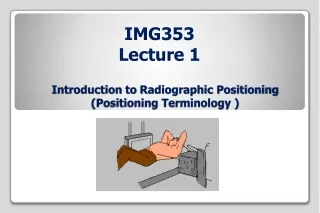
Introduction of Radiographic Technology. Radiographic Terminology Basic Imaging Principles Positioning Principles Digital Imaging. I. Radiographic Terminology. General Terms Radiograph Radiography Radiograph vs. x-ray film Radiographic images Radiographic examination or

E N D
Introduction of Radiographic Technology Radiographic Terminology Basic Imaging Principles Positioning Principles Digital Imaging
I. Radiographic Terminology • General Terms • Radiograph • Radiography • Radiograph vs. x-ray film • Radiographic images • Radiographic examination or procedure • Anatomic position
I. Radiographic Terminology • Body Plane、Section and Lines • Sagittal plane • Coronal plane • Horizontal plane • Oblique plane • Base plane • Occlusal plane
I. Radiographic Terminology • Body Surfaces and Parts • For the body • anterior • posterior • For the hands and feet • plantar • palmar • dorsum
I. Radiographic Terminology • General Body Positions • Supine • Prone • Erect (stand or sit) • Recumbent Lying down in any position • Dorsal (supine) • Ventral (prone) • Lateral
I. Radiographic Terminology • General Body Positions • Trendelenburg • Sim’s position • Fowler’s position • Lithotomy position
I. Radiographic Terminology • Specific Body Positions The body part closest to the IR (oblique and lateral) or by the surface on which the patient is lying • Lateral • Right/Left • Oblique • LPO/RPO • LAO/RAO
I. Radiographic Terminology • Decubitus (Lie on a horizontal surface and always used with horizontal x-ray beam)
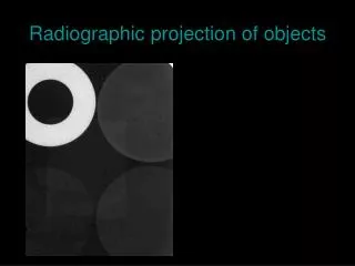
I. Radiographic Terminology • Radiographic Projection The direction or path of the CR of the x-ray beam • Anteroposterior • Posteroanterior • AP or PA Oblique • Mediolateral or Lateromedial
I. Radiographic Terminology • Additional Special Use Projection Terms • Axial • Superoinferior axial • Inferosuperior axial • AP/PA axial
I. Radiographic Terminology • Tangential • AP axial (Lordotic) • Transthoracic lateral
I. Radiographic Terminology • Dorsoplantar / Plantodorsal • Parietoacnthial / Acanthioparietal • Submentovertex /Verticosubmental
I. Radiographic Terminology • Relationship Terms • Meidal vs. Lateral • Proximal vs. Distal • Cephalad vs.Caudad
I. Radiographic Terminology • Terms Related to Movements • Flexion/Extension/Hyperextension • Ulnar deviation/Radial deviation • Dorsiflexion/Plantar flexion of foot
I. Radiographic Terminology • Terms Related to Movements • Eversion (Valgus)/Inversion(Varus) • Medial /Lateral Rotation
I. Radiographic Terminology • Terms Related to Movements • Abduction/Adduction • Supination/Pronation • Protraction/Retration
I. Radiographic Terminology • Terms Related to Movements • Elevation/Depression • Circumduction • Tilt/Rotation
I. Radiographic Terminology • Summary of Potentially Misused Terms • Position restricted to the discussion of the patient’s physical position • Projection restricted to the discussion of the path of the central ray • View restricted to the discussion of the a radiograph or image
II. Basic Imaging Principles 3 • Radiographic Criteria • Structures Show(1~6) • Position • Collimator and CR • Exposure Criteria • Image Markers a 2 1 6 c 4 b 5
II. Basic Imaging Principles • Image Markers and Patient Identification • Patient ID and Date • Anatomic side marker • Additional markers or Identification
II. Basic Imaging Principles • Radiographic Technique and Image Quality • Exposure factors • kVp • mA • S (excepted when AEC is used ) • Image Quality Factors • Density • Contrast • Detail • Distortion
II. Basic Imaging Principles • Density • Definition:the amount of blackening of the processed image • Controlling factor:mAs / kVp / SID • Change rule :Underexposure Doubling mAs • Exception:DR and CR (controlled by image process technique)
II. Basic Imaging Principles • Contrast • Definition:the difference in density on adjacent areas of a radiographic image • Purpose :make the anatomic detail of a radiographic image more visible • Controlling factor:kVp (15% increase as mAs double)
II. Basic Imaging Principles • Detail • Definition:the visible sharpness of structure on the image • Controlling factor • Geometric factors : focal spot size/SID/OID • Film/Screen Speed • Motion
II. Basic Imaging Principles • Distortion • Definition:the misrepresentation of object size or shape as projected onto film (because of beam divergence and SID)
II. Basic Imaging Principles • Distortion • Controlling factor • SID • OID/Focal spot size
II. Basic Imaging Principles • Alignment (object、film、CR)
II. Basic Imaging Principles • Anode Heel Effect Intensity of cathode > anode • Pronounced at • Shorter SID • Larger IR • Small focal spot
III. Positioning Principles • Professional Ethics and Patient Care • CAMRT (1997.06) Canadian Association of Medical Radiation Technologists • ASRT (1994.07) American Society of Radiological Technologists • Protocol and Order for General Diagnostic Radiographic Procedures • Room and Exam Preparation
III. Positioning Principles • Positioning Method • Fixed vs. Floating tabletop • Cassette tray and Bucky grid • Beam restricting device • Illuminated adjustable collimator • Positive Beam Limitation (PBL)
III. Positioning Principles • Positioning Sequences • Traditional Radiography Step1 Step2 Step3 Step4
Table • With bucky • no bucky • Standing bucky
III. Positioning Principles • Essential Projections • Routine (Basic) Projections • Commonly taken on all patients who can cooperate fully • Special (Alternate) Projections • Better demonstrate specific anatomic or certain pathology • The patients who can’t cooperate fully
III. Positioning Principles • Principle for Determining Positioning Routine • A minimum of two projections • Problem of anatomic structures being superimposed • Localization of lesions or foreign bodies • Determination of alignment of fracture • A minimum of three projections • Skeletal system involving joints • AP、PA、Oblique
III. Positioning Principles • Topographic Positioning Landmarks • Done gently • Patient should be informed • Body Habitus
III. Positioning Principles • Viewing Medical Images • Radiographic Images • AP/PA/Oblique • Viewing as patient is facing the viewer • Marked by R/L
III. Positioning Principles • Lateral • Viewing from the same perspective as the x-ray tube • Marked R/L by the side of the patient closet to the IR • Decubitus chests and abdomen • Viewing from the same perspective as the x-ray tube • Crosswise and p’t upside on view box upside • Upper/lower limb • R/L marker appears right-side-up • Limbs hanging down • Digits up
III. Positioning Principles • CT or MRI Images • The patient’s right is to the viewer’s left
IV. Digital Imaging • PACS (Picture Archiving and Communication System)
IV. Digital Imaging • CR (Computed Radiography) • Key components • Image plate (repeatedly) • IP reader (laser scanner , 20s) • Workstation • Exposure factor( AEC is not used) • Compensation 500% overexposure, 80% underexposure • Positioning consideration • Center sampling technique • Accurate and close collimation • Lead masking for multiple images • Grid
IV. Digital Imaging • DR (Digital Radiography ) • Flat panel receptor (direct conversion method ) • Digital Bucky grid 17”*17” • Automatic Exposure Control (AEC) • kVp”、”m A” manual • “s” auto
IV. Digital Imaging • DR in CGMH • Digital image unit
Quality Control for Processor • Set up initial standard (base line) • In most stable condition of the processor • Individual O.D. of 21 steps (average of five days measurements) • Find O.D. ≧1.2 Mid-density (MD) and Mid-step# • Find O.D. ≧2.2 High-density (HD) and High-step# • Find O.D. ≧0.45 Low-density (LD) and Low-step# • Daily QA • MD < ±0.15 (measurement - base line) • DD < ±0.15 (measurement - base line) (DD=HD-LD)