HUMAN ANATOMY & PHYSIOLOGY II
440 likes | 652 Vues
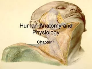
BIOL 2020. HUMAN ANATOMY & PHYSIOLOGY II. Dr. Tyler Evans Email: tyler.evans@csueastbay.edu Office: S Sci 350 Office Hours: F 8:30-11:30 or by appointment Website: http ://evanslabcsueb.weebly.com / Phone: 510-885-3475. LAST LECTURE. CHAPTER 5. t hese are all properties of your skin

HUMAN ANATOMY & PHYSIOLOGY II
E N D
Presentation Transcript
BIOL 2020 HUMAN ANATOMY & PHYSIOLOGY II Dr. Tyler Evans Email: tyler.evans@csueastbay.edu Office: S Sci 350 Office Hours: F 8:30-11:30 or by appointment Website: http://evanslabcsueb.weebly.com/ Phone: 510-885-3475
LAST LECTURE CHAPTER 5 • these are all properties of your skin • the skin and its associated components including sweat and oil glands, hair and nails make up a complex system of organs called the INTEGUMENTARY SYSTEM Fig 5.1 pg 151
LAST LECTURE CHAPTER 5 LAYERS OF THE EPIDERMIS AND DERMIS stratum corneum stratum granulosum stratum spinosum stratum basale Fig 5.2 pg 153
LAST LECTURE SKIN COLOR • three pigments contribute to skin color: MELANIN, CAROTENE and HEMOGLOBIN • only melanin is actually made in the skin • melanin is a polymer of the amino acid TYROSINE and its formation is catalyzed by the enzyme TYROSINASE Granules of melanin pigment called MELANOSOMES accumulate in the layers of skin above the dermis
LAST LECTURE APPENDAGES OF THE SKIN • skin appendages are derivatives of the epidermis and include: • Hair and hair follicles • Nails • Sweat glands • Sebaceous (oil) glands Fig 5.1 pg 151
LAST LECTURE SEVEN FUNCTIONS OF THE INTEGUMENTARY SYSTEM CHEMICAL BARRIER PHYSICAL BARRIER BODY TEMPERATURE REGULATION CUTANEOUS SENSATION METABOLIC FUNCTIONS BLOOD RESERVOIR EXCRETION
TODAY’S LECTURE: FUNDAMENTALS OF THE NERVOUS SYSTEM AND NERVOUS TISSUE CHAPTER 11
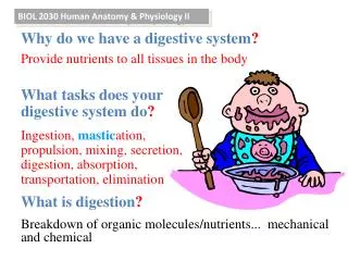
THE NERVOUS SYSTEM • the nervous system is the master controlling and communication system of the body • the nervous system has three overlapping functions: • 1. SENSORY INPUT: uses millions of receptors to monitor changes both inside and outside the body • 2. INTEGRATION: processes and interprets sensory input • 3. MOTOR OUTPUT: activates organs (e.g. muscle and glands) to appropriately respond Fig 11.1 pg 387
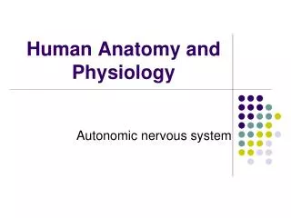
THE NERVOUS SYSTEM ORGANIZATION • We have only one highly integrated nervous system, but it is divided into several parts with specific functions: • CENTRAL NERVOUS SYSTEM (CNS): consists of the brain and spinal cord and is the integrating and control center for the nervous system • PERIPHERAL NERVOUS SYSTEM (PNS): links body parts to the CNS and consists mainly of nerves that extend out from the brain and spinal cord • PNS has two subdivisions: • a. SENSORY DIVISION: conveys nerve signals to the CNS from receptors in the body • b. MOTOR DIVISION: transmits nerve signals from the CNS to muscles and glands. • i. SOMATIC NERVOUS SYSTEM: connects to skeletal muscles • ii. AUTONOMIC NERVOUS SYSTEM: connects to smooth muscles, cardiac muscles, and glands • SYMPATHETIC DIVISION: stimulates systems • PARASYMPATHETIC DIVISION: conserves energy
THE NERVOUS SYSTEM CELL TYPES • although highly complex, the nervous system is made of two principal cell types: • 1. NEURONS: excitable cells capable of transmitting electrical signals • 2. NEUROGLIA: supporting cells that surround more delicate neurons TYPES OF NEUROGLIA: • ASTROCYTES: support, brace and anchor neurons • called SATELLITE CELLS in the PNS • MICROGLIA: repair damages neurons and prevent against infection • EPENDYMAL CELLS: line cavities of the central nervous system separating cerebrospinal fluid from nervous tissue • can be ciliated to help circulate the fluid • OLIGODENDROCYTES: wrap around neurons to produce an insulating cover called MYELIN SHEATH • in the PNS, SCHWANN CELLS produce myelin sheath Fig 11.3 pg 390
THE NERVOUS SYSTEM TYPES OF NEURONS • neurons are the structural units of the CNS: they have extreme longevity (entire lifetime), once formed they do not divide and required a large amount of energy • while neurons come in many cell types, they share common features • e.g. vertebrate motor neuron
THE NERVOUS SYSTEM TYPES OF NEURONS • INTERNEURONS connect other neurons of the CNS • can lack a clearly defined axon because are only transmitting signals over short distances between other neurons. • SENSORY NEURONS transmit signals from receptors on skin or internal organs to the CNS • EFFERENT or MOTOR NEURONS: carry impulses away from the CNS to muscles and glands • long axons of sensory and efferent neurons help transmit signals over greater distances
THE NERVOUS SYSTEM TYPES OF NEURONS • although most neurons have the same basic components, each of these components has been modified best perform specific tasks • structure dictates function • most neurons have DENDRITES, a CELL BODY (SOMA) and an AXON, but details of each structure are variable • this diversity allows neurons to perform different tasks in the nervous system
THE NERVOUS SYSTEM MYELIN SHEATH • many axons, particularly long or thick ones, are covered with a whitish and fatty MYELIN SHEATH • protects and electrically insulates axons (i.e. prevents signals from decaying) and increases the speed of signal transmission down axons • SCHWANN CELLS wrap themselves around the axon many times and then squeeze the cytoplasm out so that just the plasma membrane remains • gaps between individual Schwann cells form NODES OF RANVIER • in the CNS, OLIGODENDROCYTES form the myelin sheath, but use a similar process • regions of the brain that contain a lot of myelinated axons appear white and is called WHITE MATTER • unmyelinated axons from GREY MATTER Fig 11.5 pg 392
THE NERVOUS SYSTEM MEMBRANE POTENTIALS • when a neuron is activated, an electrical signal called a POTENTIAL travels down its axon, • these signals underlie virtually every function in the nervous system • recall, that integral proteins in cell membranes called ION CHANNEL PROTEINS control the movement of charged particles across the membrane and this generates an ELECTROCHEMICAL GRADIENT. • when channel proteins open, ions move down their concentration gradient creating an electrical charge • changes in membrane potential produce two types of electrical signals: • 1. GRADED POTENTIALS: weak and operate over a short distances • 2. ACTION POTENTIALS: strong and operate over longer distances
THE NERVOUS SYSTEM ACTION POTENTIALS • only neurons and muscle cells can generate action potentials (AP) • an AP is a brief reversal of membrane charge (from about -70 mV to +30 mV) • unlike graded potentials, action potentials do not degrade over distance • AP are created by the movements of sodium (Na+) and potassium (K+) across the membrane via integral proteins. The sequence of events are as follows: • RESTING STATE: Na+ and K+ protein channels are closed • DEPOLARIZATION: Na+ channels open and Na+ rushes into cells, which triggers the opening of even more Na+ channels in a POSITIVE FEEDBACK MECHANISM • REPOLARIZATION: Na+ channels close and K+ channels open and K+ rushes out of cells, which works to restore the resting membrane potential • HYPERPOLARIZATION: K+ remain open for longer than required and excessive K+ moves out of cells causing a temporary drop below the resting state • resting state is returned as Na+ channels close • Na+, K+ ATPase then works to restore the correct number of Na+ and K+ ions to each side of the membrane
THE NERVOUS SYSTEM ACTION POTENTIALS Changes in potential Changes in permeability (i.e. opening or closing of ion channels) Fig 11.11 pg 402-403
THE NERVOUS SYSTEM CONDUCTION VELOCITY • conduction velocities vary between neurons: • larger diameter axons propagate signals faster than smaller ones • myelinated axons propagate signals faster than non-myelinated axons • myelination prevents signal from spreading onto adjacent cell membranes and the signal confined only to the single axon • In MULTIPLE SCLEROSIS, the body’s immune system destroys myelin sheath and the signal degrades and impairs movement Fig 11.15 pg 406
THE NERVOUS SYSTEM THE SYNAPSE • aSYNAPSE is the junction that connects one neuron to another or to an effector organ like a muscle or gland • synapses occur in two varieties: • ELECTRICAL SYNAPSES: are less common and occur via proteins connections called CONNEXONS that connect the cytoplasm of adjacent cells and allow the movement of small molecules between cells • conduction across electrical synapses is very fast
THE NERVOUS SYSTEM SYNAPTIC TRANSMISSION • synapses occur in two varieties: • 2. CHEMICAL SYNAPSES: are specialized to allow the release and reception of NEUROTRANSMITTERS, a large group of chemicals that exert a variety of effects on the body • chemical synapses consists of a AXON TERMINAL on the pre-synaptic neuron which are packed with synaptic vesicles that contain neurotransmitters and a RECEPTOR REGION on the post-synaptic neuron. • these structures are separated by a gap called the SYNPATIC CLEFT • when an AP arrives at the axon terminal, it triggers calcium (Ca+2) channels to open. Ca+2 rushes into the cell where it acts as a signal for synaptic vesicles to unload their neurotransmitters into the cleft • neurotransmitters diffuses across the cleft and binds to receptors on integral membrane proteins of the post-synaptic neuron. • different types of neurotransmitter effect different channel proteins and therefore result in different outcomes either EXCITATORY or INHIBITORY
THE NERVOUS SYSTEM SYNAPTIC TRANSMISSION Fig 11.17 pg 409
THE CENTRAL NERVOUS SYSTEM CHAPTER 12: THE BRAIN • recall, CNS consists of the BRAIN and SPINAL CORD and is the integrating and control center for the nervous system • four regions in the adult brain: • CEREBRAL HEMISPHERES • DIENCEPHALON • BRAIN STEM • CEREBELLUM (includes midbrain, pons and medulla oblongata) Fig 12.2 pg 430
THE CENTRAL NERVOUS SYSTEM THE BRAIN • the CEREBRAL HEMISPHERES form the most conspicuous parts of the brain, together accounting for 83% of the total mass • elevated ridges are called GYRI, which are separated by grooves called SULCI • several sulci divide each hemisphere into five LOBES: • FRONTAL LOBE: occurs at the anterior (front) of the skull • PARIETAL LOBE • TEMPORAL LOBE • OCCIPITAL LOBE: occurs at the posterior (back) of the skull • INSULA: buried underneath frontal and temporal lobes Fig 12.4 pg 432
THE CENTRAL NERVOUS SYSTEM THE BRAIN • the CEREBRAL CORTEX is where the conscious mind is found and allows communication, memory and learning • composed of GREY MATTER and accounts for 40% of the brain’s mass Fig 12.4 pg 432
THE CENTRAL NERVOUS SYSTEM THE BRAIN • the CEREBRAL CORTEX is where the conscious mind is found and allows communication, memory and learning • composed of GREY MATTER and accounts for 40% of the brain’s mass Fig 12.4 pg 432
THE CENTRAL NERVOUS SYSTEM THE BRAIN • the CEREBRAL CORTEX is where the conscious mind is found and allows communication, memory and learning • composed of GREY MATTER and accounts for 40% of the brain’s mass Fig 12.4 pg 432 • the cerebral cortex is divided into three functional areas (actually a simplification as not each area acts entirely alone): • MOTOR AREAS: control voluntary movement • SENSORY AREAS: concerned with the conscious awareness of sensation • ASSOCIATION AREAS: where individual perceptions are integrated into a more unified process
THE CENTRAL NERVOUS SYSTEM MOTOR AREAS • MOTOR AREAS of the cerebral cortex lie in the posterior sections of the frontal lobes and comprise four areas: • PRIMARY MOTOR CORTEX: large neurons called PYRIMIDAL CELLSallow this region to control the movement of skeletal muscles • sub-regions of the motor cortex control specific body areas (e.g. feet, arms, etc.) • PRE-MOTOR CORTEX: coordinates the movement of several muscle groups into complex tasks (e.g. playing a musical instrument) • BROCA’S AREA: directs muscles involved in speech production • FRONTAL EYE FIELD: controls the voluntary movement of the eyes
THE CENTRAL NERVOUS SYSTEM MOTOR AREAS • MOTOR AREAS of the cerebral cortex lie in the posterior sections of the frontal lobes and comprise four areas: Fig 12.6 pg 434
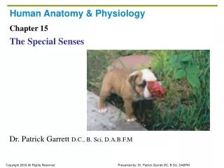
THE CENTRAL NERVOUS SYSTEM SENSORY AREAS • SENSORY AREAS concerned with the conscious awareness of sensation and comprise eight areas: • PRIMARY SOMATOSENSORY CORTEX: receive input from touch SOMATOSENSORY ASSOCIATION CORTEX: integrate sensory information to understand an object being felt • VISUAL AREAS: largest sensory area and receives input from the retina • AUDITORY AREAS: detect sound energy exiting inner ear • VESTIBULAR CORTEX: conscious awareness of balance • OLFACTORY CORTEX: is connected to odor receptors in nose via long axons and is involved in smell • GUSTATORY CORTEX: region involved in taste • VISCERAL SENSORY AREA: detects sensations like an upset stomach, full bladder and need for air when holding breath
THE CENTRAL NERVOUS SYSTEM SENSORY AREAS • SENSORY AREAS concerned with the conscious awareness of sensation and comprise eight areas: Fig 12.6 pg 434
THE CENTRAL NERVOUS SYSTEM ASSOCIATION AREAS • ASSOCIATION AREAS where individual perceptions are integrated into a more unified process • gives meaning to the information we receive, allows us to store it memory, and decide what action to take • damage to the association area impairs judgment and causes a loss of inhibitions • also cause failure to recognize parts of their body as belonging to themselves
THE CENTRAL NERVOUS SYSTEM WHITE MATTER • WHITE MATTER underlies grey matter and is responsible for communication within the CNS and contain three nerve fiber types: • ASSOCIATION FIBERS: connect different parts of the same hemisphere • COMMISSURAL FIBERS: allows both hemispheres to function as a whole • PROJECTION FIBERS: processes sensory information Fig 12.8 pg 438
THE CENTRAL NERVOUS SYSTEM DIENCEPHALON • up until now we have been describing parts of the cerebral hemispheres • the DIENCEPHALON is the next major brain region and lies at the core of the forebrain. It consists largely of three paired structures: • THALAMUS: sensory information converges here information is sorted out and edited (i.e. what is important) • HYPOTHALAMUS: controls the viscera (internal organs) and is very important in maintaining homeostasis, such as body temperature, food intake and thirst. • EPITHALAMUS: regulates sleep/wake cycles Fig 12.8 pg 440
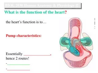
THE CENTRAL NERVOUS SYSTEM BRAIN STEM • the brain stem produces the programmed actions required for survival. • also associated with 10 of the 12 pairs of cranial nerves. • there are three brain stem regions: • MIDBRAIN: functions associated with pain suppression, fight or flight response and hearing • PONS: composed of nerve fibers • MEDULLA OBLONGATA: crucial role as an autonomic reflex center, including • a. CARDIOVASCULAR CENTER: adjusts heart contraction force and rate • b. RESPIRATORY CENTER: generates respiratory rhythm • c. OTHER CENTERS: vomiting, hiccuping, swallowing, coughing and sneezing Fig 12.13 pg 444
THE CENTRAL NERVOUS SYSTEM BRAIN STEM-FACIAL NERVES (FOR LAB ONLY) Fig 12.13 pg 444
THE CENTRAL NERVOUS SYSTEM CEREBELLUM • the CEREBELLUM provides the precise timing and appropriate patterns of skeletal muscle contraction for smooth movement (e.g. driving, typing, etc) • activity occurs subconsciously, we are not aware of it • damage to the cerebellum results in clumsy unsure movements Fig 12.15 pg 448
THE CENTRAL NERVOUS SYSTEM LIMBIC SYSTEM • refers to a group of structure located on the medial aspect of each hemisphere • limbic system is our emotional brain, especially the AMYGDALOID BODY (involved in fear responses) and CINGULATE GYRUS (involved in facial expressions) Fig 12.16 pg 449
THE CENTRAL NERVOUS SYSTEM BRAIN PATHOLOGIES • single most common nervous system disorder and third leading cause of death in the U.S. are CEREBROVASCULAR ACCIDENTS, also called STROKES • occurs when blood circulation to the brain is blocked and brain tissue dies • mostly commonly a blot clot that blocks an artery to the brain. Clots form on the walls of arteries narrowed by ATHEROSCLEROSIS • those affected can be partially paralyzed and have difficulty with speech
THE CENTRAL NERVOUS SYSTEM BRAIN PATHOLOGIES • up to 50% of people over 85 years of age have ALZHEIMER’S DISEASE • memory loss, shortened life span, disorientation, confusion and irritability • caused by accumulations of BETA-AMYLOID PEPTIDES that disrupt brain function
THE CENTRAL NERVOUS SYSTEM THE SPINAL CORD • the spinal cord is enclosed in the vertebral column and provides a two-way conduction pathway to/from the brain • surrounded by a protective membrane called DURA MATER and CEREBROSPINAL FLUID • 31 pairs of spinal nerve fibers attach via foramen (holes) in vertebrae • these fibers connect to motor and sensory neurons that control movement and the senses Fig 12.28 pg 467 Fig 12.26 pg 465
THE CENTRAL NERVOUS SYSTEM SPINAL CORD TRAUMA • damage to the spinal cord results in PARALYSIS (loss of motor function) or PARATHESIAS (abnormal sensations) • severing of the spinal cord results in total motor and sensory loss in body regions inferior (below) this site of damage • if damage occurs in thoracic or lumbar regions both lower limbs are impaired called PARAPLEGIA • if damage occurs in the cervical section all four limbs are affected and this is referred to as QUADRIPLEGIA • POLIOMYELITIS inflammation of the spinal cord from ingesting water contaminated with poliovirus. The virus destroys neurons of the spinal cord leading to motor impairment and paralysis • eradicated with vaccines, but infection rates increasing as parents choose not to vaccinate their children • AMYOTROPHIC LATERAL SCLEROSIS: neuromuscular conditions caused by destruction of neurons of the spinal cord and patient loses ability to speak, swallow and breathe • also called Lou Gehrig’s disease
FOR REVIEW TONIGHT • understand the organization of the nervous system • understand the structure and function of nerve cells • understand how action potentials are generated and the events occurring at the synapse • understand the structure and function of the four major regions of the brain (i.e. CEREBRAL HEMISPHERES, DIENCEPHALON, BRAIN STEM, CEREBELLUM)
NEXT LECTURE SPECIAL SENSES CHAPTER 15