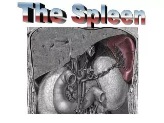
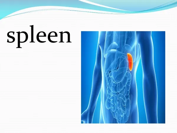
spleen
spleen. Clinical Applied Anatomy Embryology: Dorsal Mesogastrium. The weight is 75-250 g. Anatomy: Lt Hypochondrial region.( 11-9 th rib) along the 10 th rib upto Mid Ax. Line.

spleen
E N D
Presentation Transcript
Clinical Applied Anatomy • Embryology: Dorsal Mesogastrium. • The weight is 75-250 g. • Anatomy: Lt Hypochondrial region.( 11-9th rib) along the 10 th rib upto Mid Ax. Line. • The notch is in the I.L. border may be papable. the retroperitoneal attachments of the spleen, the splenocolic, splenorenal, and splenophrenic ligaments - Splenic. Arteryruns along the upper border of PancreasAnd is divided into Sup. & Inf. Branches.
- Splenic Vein join superior mescentric vein to form portal vein. • - Lymphatic drainage by lymph vessels of the white pulp. • - Accessory Spleen ( Splenunculi ) in hilum most common, pedicle, tail of pancreas, Sp-Colic lig., G.O. & Mesentry.
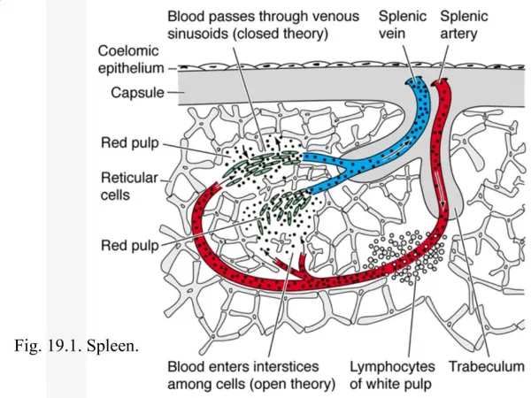
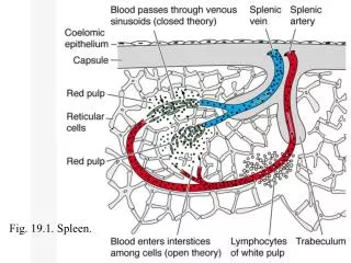
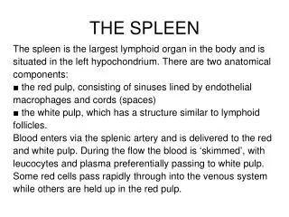
Physiology and histology: The spleen consist of trabicular vessels surrounded by the white pulp, then marginal zone which is covered by red pulp. The white pulp consist of lymphatic sheaths contains plasma cells (Igs), and T lymphocytes. It represent the immune center of the spleen and 25% of its weight. The red pulp consist of cords and sinuses and represent the hemopoietic function of the spleen.
Functions of Spleen • Immune Function. IgM. Production and foreign antigens processing. • Filter Function. ( Cellular & Non Cellular ). • Pitting. Repair of RBC.( Inclusion bodies). • Resevoir . • Cytopoiesis.
Assessment • History, Examination, Investigations • Palpation Technique Start in RLQ (so you don’t miss a giant spleen). Get your fingers set then ask patient to take a deep breath. Don’t dip your fingers or do anything but wait.When patient expires, take up new position. Note lowest point of spleen below costal margin, texture of splenic contour, and tenderness If spleen is not felt, repeat with pt lying on right side. Gravity may bring spleen within reach.
“LET THE SPLEEN PALPATE YOUR FINGERS AND NOT THE OTHER WAY AROUND. THERE IS NO GOLD, SO DON’T DIG!” • Remember that the spleen can become very enlarged and fragile (e.g. infectious mononucleosis); overly aggressive palpation may cause injury
Investigations • Laboratories. • Imaging. Plain XR, U.S, Doppler, CT scan, MRI & 99mTc – labelled colloid. • .splenectomy
Diseases • Trauma • Isolated • or Combined Disorders of Spleen: • Congenital abnormality • Blunt • Splenic artery aneurysm, infarct & rupture. • Penetrated • Splenomegaly & Hyperspleenism. • Neoplasms
Congenital Abnormality Splenic agenesis 10% in children with CHD. Polysplenia is rare due to failure of splenic fusion. They are functionless. 3. Splenunculi : Accessory spleens, 10 – 30% of population, hilum 50%, tail of pancreas 30%, mesocolon, splenic ligament & G.O. They are functioning. You have to identify them during splenectomy.
Hamartomas Splenic Hamartomas is a rare, benign vascular proliferation that is often found incidentally while working up on other complaints or at autopsy. Women more commonly present with symptoms related to mass effect than men. Histologically : it may resemble the white or red pulp.Treatment: Splenectomy for Diagnostic purpose to exclude malignancy or for pressure symptoms.
Non paracytic Cyst • Primary Cyst ( True ) • Secondary (pseudocyst) caused by trauma. • True wall ,may be calcified. • ( Cystic haemangioma,lymphangiomas and dermoid and epidermoid cyst). • DDx is psuedocyst.& hydatid cyst. • Treatment: if > 5 cm Splenectomy.
Splenic Abscess Infected embolus, association or with typhoid & PT fever, osteomyelitis, otitis media, infected splenic infarct, pancreatic necrosis or other intra abdominal infection & puerperal sepsis. Complication : 1. Lt sub diaphragmatic abscess 2. peritonitis Treatment: is of underlying cause, plus either: Percutaneous Drainage under U/S guidance if unilocular or Surgical drainage if multilocular.
Splenic Rupture • Due to blunt abdominal trauma to the Lt UQ of the abdomen. • Emergency case . • DDx: Rupture haemangioma, Rupture hydatid cyst., Rupture malarial spleen. Ruptured malarial spleen. Is the most common cause of non traumatic rupture.
Splenic artery aneurysm • Occur in 0.04-1 per cent of population. • Twice more common in females. • Usually single, multiple in a quarter of cases. • Caused by : intra-abdominal sepsis, pancreatic necrosis and atherosclerosis. • Its usually symptomless but present as rupture. • Surgery is indicated in the following cases: • 1.Symptomatic • 2.More than 2cm • Pregnancy • pancreatitis • .
Surgical intervention includes: Embolization Endovascular stenting Ligation of proximal and distal ends of the sac Partial or complete appendectomy
Splenomegaly Hypersplenism Clinical syndrome that is characterized by splenic enlargement and any combination of anaemia, leukopenia or thrombocytopenia, compensatory B.M. hyperplasia & improvement after splenectomy.
INFECTIONBacterialT.B., Abscess. Viral HIV related TCP Protozoa & Parasitic Schistosomiasis H.C. Tropical Splenomegaly BLOOD DISEASE ITP Hereditary Spherocytosis Autoimmune Haemolytic Anaemia Thalassemia Sickle Cell Disease METABOLIC Gaucher’s disease, Porphyria CIRCULATORYSegmental Portal HT COLLAGEN disease Felty’s syndrome NEOPLASTIC Hodjkin Lymphoma mylofibrosis
Tuberculosis Cardinal symptoms is: • Young age with Splenomegaly, asthenia weight loss and fever. it may cause portal HT rarely cold abscess . Treatment :anti TB drugs . Splenectomy is not normally required Tropical SPLENOMEGALY Massive splenomegally caused by Malaria, Kala-azar & Schistosomiasis. . May require splenectomy for anaemia or pressure symptoms. Antimalaria drugs may be indicated for long life in endemic areas.
Schistosomiasis: • Its prevalent in Africa, Asia, and South America . • Schistosoma Mansoni in 75% of cases. • Splenomegally result from portal hypertension. • Splenectomy indicated for symptomatic relief.
Idiopathic thrombocytopenic purpura • Low platelet count result from the development of IgG. • Associated with normal bone marrow and absence of other causes of thrombocytopenia. • It can be divided into child and adult form.
ITP CHILD FORM ADULT FORM • Acute • Follows acute infection • Spontaneous resolution within 2 months • Occur in 50 cases per million annually. • Chronic • No specific cause • Persist longer than six months • 50-75 cases per million
Spleen is palpable in 10% of cases. • Intracranial hemorrhage is the most common cause of death. • Treatment: • Started if platelet fall below 10,000, includes • Steroids • Intravenous Igs. • splenectomy, indicated if • 2 relapse on steroid therapy • Platelet count remains low • Treatment persist 6-9 months.
HERIDITARY SPHEROCYTOSIS • Autosomal dominant • RBCs are spherocytic • There is defect in membrane proteins: alpha and beta spectrin, ankyrin, band 3 protein and protein 4.2. • There is increase membrane permeability to Na, influx of Na into the cell increase osmotic pressure resulting in swelling and fragility of the RBCs. • Why splenomegally occur?
Clinical features: Usually present in childhood Anaemia Jaundice Gallstone Splenomegally, may be hepatomegally Leg ulcers Diagnosis: Blood film: round shape RBCs Fragility test: RBCs hemolyse in 0.6% solution Increase faecal urobilinogen Radioactive chromium
Treatment: All patients need splenectomy, in juvenile cases it should be postponded to the age of 6 years but before the development of gallstones.
Acquired autoimmune haemolytic anaemia • It may arise following exposure to chemicals, infection or drugs e.g. alpha-methyldopa. • More common in females. • Splenomegally occur in 50% • Gallstone occur in 20% • Divided into immune (diagnosed by coombes test) and non immune • Treatment: steroid, if failed or complication develop then proceed to splenectomy.
Thalassemia • Transmitted as recessive trait • Divided into alpha, beta and gamma • Most patients suffers from beta(major) type. • Diagnosis: by 1. resistance to osmotic lysis • 2. Hb electrophoresis • Treatment: • Blood transfusion • Splenectomy: in transfusion dependent patient and if haemolytic antibodies develop.