THE SPLEEN
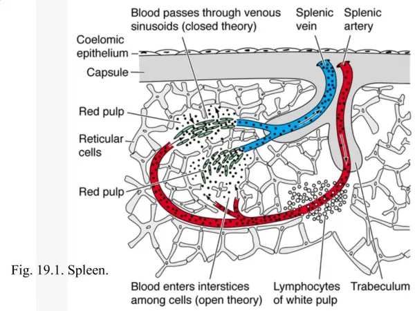
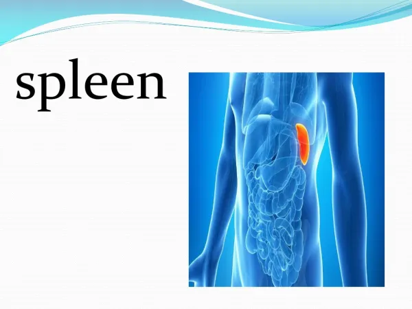
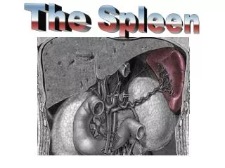
THE SPLEEN. The spleen is the largest lymphoid organ in the body and is situated in the left hypochondrium. There are two anatomical components: ■ the red pulp, consisting of sinuses lined by endothelial macrophages and cords (spaces)

THE SPLEEN
E N D
Presentation Transcript
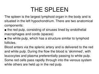
THE SPLEEN The spleen is the largest lymphoid organ in the body and is situated in the left hypochondrium. There are two anatomical components: ■ the red pulp, consisting of sinuses lined by endothelial macrophages and cords (spaces) ■ the white pulp, which has a structure similar to lymphoid follicles. Blood enters via the splenic artery and is delivered to the red and white pulp. During the flow the blood is ‘skimmed’, with leucocytes and plasma preferentially passing to white pulp. Some red cells pass rapidly through into the venous system while others are held up in the red pulp.
Functions Sequestration and phagocytosis. Normal red cells, which are flexible, pass through the red pulp into the venous system without difficulty. Old or abnormal cells are damaged by the hypoxia, low glucose and low pH found in the sinuses of the red pulp and are therefore removed by phagocytosis along with other circulating foreign matter. Howell–Jolly and Heinz bodies and sideroblastic granules have their particles removed by ‘pitting’ and are then returned to the circulation. IgG-coated red cells are removed through their Fc receptors by macrophages.
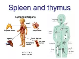
Extramedullary haemopoiesis. Pluripotential stem cells are present in the spleen and proliferate during severe haematological stress, such as in haemolytic anaemia or thalassaemia major. Immunological function. About 25% of the body’s T lymphocytes and 15% of B lymphocytes are present in the spleen. The spleen shares the function of production of antibodies with other lymphoid tissues. Blood pooling. Up to one-third of the platelets are sequestrated in the spleen and can be rapidly mobilized. Enlarged spleens pool a significant percentage (up to 40%) of the red cell mass.
SPLENOMEGALY Causes A clinically palpable spleen can have many causes. ■ Infection: (a) acute, e.g. septic shock, infective endocarditis, typhoid, infectious mononucleosis (b) chronic, e.g. tuberculosis and brucellosis (c) parasitic, e.g. malaria, kala-azar and schistosomiasis. ■ Inflammation: rheumatoid arthritis, sarcoidosis, SLE. ■ Haematological: haemolytic anaemia, haemoglobinopathies and the leukaemias, lymphomas and myeloproliferative disorders. ■ Portal hypertension: liver disease. ■ Miscellaneous: storage diseases, amyloid, primary and secondary neoplasias, tropical splenomegaly Massive splenomegaly. This is seen in myelofibrosis, chronic myeloid leukaemia, chronic malaria, kala-azar or, rarely, Gaucher’s disease.
Hypersplenism This can result from splenomegaly due to any cause. It is commonly seen with splenomegaly due to haematological disorders, portal hypertension, rheumatoid arthritis (Felty’s syndrome) and lymphoma. Hypersplenism produces: ■ pancytopenia ■ haemolysis due to sequestration and destruction of red cells in the spleen ■ increased plasma volume. Treatment. This is often dependent on the underlying cause, but splenectomy is sometimes required for severe anaemia or thrombocytopenia
Splenectomy Splenectomy is performed mainly for: ■ trauma ■ immune thrombocytopenic purpura ■ haemolytic anaemias ■ hypersplenism.
Problems after splenectomy An immediate problem is an increased platelet count (usually 600–1000 × 109/L) for 2–3 weeks. Thrombo-embolic phenomena may occur. In the longer term there is an increased risk of overwhelming infections, particularly pneumococcal infections.
Prophylaxis against infection after splenectomy or splenic dysfunction
Vaccinate 2–3 weeks before elective splenectomy. ■ A 23-valent unconjugated pneumococcal polysaccharide vaccine repeated every 5 years ■ Meningococcal group C conjugate vaccine ■ Annual influenza vaccine ■ Haemophilus influenzae type b (Hib) vaccine ■ Long-term penicillin V 500 mg 12-hourly (if sensitive, use erythromycin) ■ Meningococcal polysaccharide vaccine (ACWY) for travellers to Africa/Saudi Arabia, e.g. during Hajj and Umrah pilgrimages
Prophylaxis against infection after splenectomy or splenic dysfunction All patients should be educated about the risk of infection and the importance of its early recognition and treatment. They should be given an information leaflet and should carry a card or bracelet to alert health professionals to their risk of overwhelming infection
Postsplenectomy haematological features ■ Thrombocytosis persists in about 30% of cases. ■ The WBC count is usually normal but there may be a mild lymphocytosis and monocytosis. ■ Abnormalities in red cell morphology are the most prominent changes and include Howell–Jolly bodies (contain basophilic nuclear remnants), Pappenheimer bodies (contain sideroblastic granules), target cells and irregular contracted red cells. Pitted red cells can be counted.
Splenic atrophy This is seen in sickle cell disease due to infarction. It is also seen in coeliac disease, in dermatitis herpetiformis, and occasionally in ulcerative colitis and essential thrombocythaemia. Postsplenectomy haematological features are seen.
The cells and proteins in the blood express antigens which are controlled by polymorphic genes; that is, a specific antigen may be present in some individuals but not in others. A blood transfusion may immunize the recipient against donor antigens that the recipient lacks (alloimmunization), and repeated transfusions increase the risk of the occurrence of alloimmunization. Similarly, the transplacental passage of fetal blood cells during pregnancy may alloimmunize the mother against fetal antigens inherited from the father. Antibodies stimulated by blood transfusion or pregnancy, such as Rh antibodies, are termed immune antibodies and are usually IgG, in contrast to naturally occurring antibodies, such as ABO antibodies, which are made in response to environmental antigens present in food and bacteria and which are usually IgM.
BLOOD GROUPS The blood groups are determined by antigens on the surface of red cells; more than 280 blood groups are recognized. The ABO and Rh systems are the two major blood groups, but incompatibilities involving many other blood groups (e.g. Kell, Duffy, Kidd) may cause haemolytic transfusion reactions and/or haemolytic disease of the newborn (HDN).
ABO system This blood group system involves naturally occurring IgM anti-A and anti-B antibodies which are capable of producing rapid and severe intravascular haemolysis of incompatible red cells. The ABO system is under the control of a pair of allelic genes, H and h, and also three allelic genes, A, B and O, producing the genotypes and phenotypes . The A, B and H antigens are very similar in structure; differences in the terminal sugars determine their specificity. The H gene codes for enzyme H, which attaches fucose to the basic glycoprotein backbone to form H substance, which is the precursor for A and B antigens).
The A and B genes control specific enzymes responsible for the addition to H substance of N-acetylgalactosamine for Group A and d-galactose for Group B. The O gene is amorphic and does not transform H substance and therefore O is not antigenic. The A, B and H antigens are present on most body cells. These antigens are also found in soluble form in tissue fluids such as saliva and gastric juice in the 80% of the population who possess secretor genes
The ABO system: antigensand antibodies Phenotype Genotype Antigens Antibodies Frequency UK (%) O OO None Anti-A and anti-B 43 A AA or AO A Anti-B 45 B BB or BO B Anti-A 9 AB AB A and B None 3
Rh system There is a high frequency of development of IgG RhD antibodies in RhD-negative individuals after exposure to RhD positive red cells. The antibodies formed cause HDN and haemolytic transfusion reactions. This system is coded by allelic genes, C and c, E and e, D and no D, which is signified as d; they are inherited as triplets on each chromosome 1, one from each pair of genes (i.e. CDE/cde). RhD-negative individuals have no D protein in the red cell membrane which explains why it is so immunogenic. In Caucasians, the RhD-negative phenotype almost always results from a complete deletion of the RhD gene; in black Africans, it can also result from an inactive gene containing stop codons in the reading frame.
PROCEDURE FOR BLOOD TRANSFUSION The safety of blood transfusion depends on meticulous attention to detail at each stage leading to and during the transfusion. Avoidance of simple errors involving patient and blood sample identification at the time of collection of the sample for compatibility testing and at the time of transfusion would avoid most serious haemolytic transfusion reactions, almost all of which involve the ABO system
Pretransfusion compatibility testing Blood grouping The ABO and RhD groups of the patient are determined. Antibody screening The patient’s serum or plasma is screened for atypical antibodies that may cause a significant reduction in the survival of the transfused red cells. The patient’s serum or plasma istested against red cells from at least two group O donors, expressing a wide range of red cell antigens, for detection of IgM red cell alloantibodies (using a direct agglutination test of cells suspended in saline) and IgG antibodies (using an indirect antiglobulin test. About 10% of patients have a positive antibody screening result; in which case, further testing is carried out using a comprehensive panel of typed red cells to determine the blood group specificity of the antibody (clinically significant red cell antibodies are detected in about 20% of patients with positive antibody screens).
Selection of donor blood and crossmatching Donor blood of the same ABO and RhD group as the patient is selected. Matching for additional blood groups is carried out for patients with clinically significant red cell antibodies for patients who are likely to be multitransfused and at high risk of developing antibodies, e.g. sickle cell disease, and many centres routinely provide c-negative and Kell-negative blood for women of child-bearing age to minimize the risk of alloimmunization and subsequent HDN
COMPLICATIONS OF BLOODTRANSFUSION Immunological Alloimmunization and incompatibility Red cells Immediate haemolytic transfusion reactions. Delayed haemolytic transfusion reactions. Leucocytes and platelets Non-haemolytic (febrile) transfusion reactions Post-transfusion purpura Poor survival of transfused platelets and granulocytes Graft-versus-host disease Lung injury (TRALI) Plasma proteins Urticarial and anaphylactic reactions
Transmission of infection Viruses – HAV, HBV, HCV – HIV, HHV8 – CMV, EBV, HTLV-1, WNV Parasites – malaria, trypanosomiasis, toxoplasmosis Bacteria Prion – CJD Circulatory failure due to volume overload Iron overload due to multiple transfusions Massive transfusion of stored blood may cause bleeding reactions and electrolyte changes Physical damage due to freezing or heating Thrombophlebitis Air emboiism
Incompatibility This may result in poor survival of transfused cells, such as red cells and platelets, and also in the harmful effects of antigen–antibody reaction
1. Red cells Haemolytic transfusion reactions Immediate reaction. This is the most serious complication of blood transfusion and is usually due to ABO incompatibility. There is complement activation by the antigen–antibody reaction, usually caused by IgM antibodies, leading to rigors, lumbar pain, dyspnoea, hypotension, haemoglobinuria and renal failure. The initial symptoms may occur a few minutes after starting the transfusion. Activation of coagulation also occurs and bleeding due to disseminated intravascular coagulation (DIC) is a bad prognostic sign. Emergency treatment for shock .is needed to maintain the blood pressure and renal function.
Diagnosis This is confirmed by finding evidence of haemolysis (e.g. haemoglobinuria), and incompatibility between donor and recipient. All documentation should be checked to detect errors such as: ■ failure to check the identity of the patient when taking the sample for compatibility testing (i.e. sample from the wrong patient) ■ mislabelling the blood sample with the wrong patient’s name ■ simple labelling or handling errors in the laboratory ■ errors in the collection of blood, leading to delivery of the wrong blood to the ward/theatre ■ failure to perform proper identity checks before the blood is transfused (i.e. blood transfused to the wrong patient).
Investigations To confirm where the error occurred, blood grouping should be carried out on: ■ the patient’s original sample (used for the compatibility testing) ■ a new sample taken from the patient after the reaction ■ the donor units. At the first suspicion of any serious transfusion reaction, the transfusion should always be stopped and the donor units returned to the blood transfusion laboratory with a new blood sample from the patient to exclude a haemolytic transfusion reaction.
Delayed reaction. This occurs in patients alloimmunized by previous transfusions or pregnancies. The antibody level is too low to be detected by pretransfusion compatibility testing, but a secondary immune response occurs after transfusion, resulting in destruction of the transfused cells, usually by IgG antibodies. Haemolysis is usually extravascular as the antibodies are IgG, and the patient may develop anaemia and jaundice about a week after the transfusion, although most of these episodes are clinically silent. The blood film shows spherocytosis and reticulocytosis. The direct antiglobulin test is positive and detection of the antibody is usually straightforward.
2. Leucocytes and platelets Non-haemolytic (febrile) transfusion reactions Febrile reactions are a common complication of blood transfusion in patients who have previously been transfused or pregnant. The usual causes are the presence of leucocyte antibodies in an alloimmunized recipient acting against donor leucocytes in red cell concentrates leading to release of pyrogens, or the release of cytokines from donor leucocytes in platelet concentrates. Typical signs are flushing and tachycardia, fever (> 38°C), chills and rigors. Aspirin may be used to reduce the fever, although it should not be used in patients with thrombocytopenia. The introduction of leucocyte depleted blood in the UK, to minimize the risk of transmission of variant Creutzfeldt–Jakob disease (vCJD) by blood transfusion , has reduced the incidence of febrile reactions.
Potent leucocyte antibodies in the plasma of donors, who are usually multiparous women, may cause severe pulmonary reactions (called transfusion-related acute lung injury or TRALI) characterized by dyspnoea, fever, cough, and shadowing in the perihilar and lower lung fields on the chest X-ray. Prompt respiratory support is essential; mechanical ventilation is frequently necessary. It usually resolves within 48–96 hours, but the mortality is approximately 20% in the 185 cases