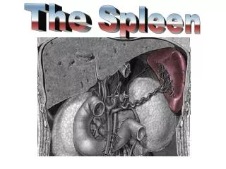
SPLEEN
This detailed overview discusses the development, anatomy, and histology of the spleen in human anatomy. Beginning with the differentiation of mesenchymal cells in the dorsal mesogastrium during the fifth week of development, it details the formation of lymphoblasts and other blood-forming cells. The anatomy highlights key features such as size, shape, and peritoneal connections, while also covering the microstructure including white and red pulp. The document further addresses congenital anomalies and accessory spleens, providing a comprehensive understanding of this vital organ.


SPLEEN
E N D
Presentation Transcript
SPLEEN ROY GEORGE SHARON
DEVELOPMENT • Collection of mesenchymal cells in the dorsal mesogastrium- 5th week • Differentiate into lymphoblasts and other blood forming cells • Mass projects to left, covered by peritoneum • Dorsal mesogastriumgastrosplenic lig. lienorenal lig.
Contd. • Fusion of lienorenal lig to post.abdominal wall • Change in orientation of stomach • Spleen lies on left side forms the left boundary of the lesser sac of peritoneum
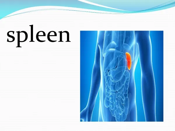
Large encapsulated mass of vascular and lymphoid tissue • SITUATION • Upper Lt. quadrant of the abdomen b/n the fundus of stomach and diaphragm • SHAPE • Slightly curved wedge to ‘domed’ tetrahedron
SIZE • 12cm long , 7cm broad , 3-4 cm wide • WEIGHT • 150 gm (80gm -250gm)
Relations of spleen • SURFACES • Superolateral • Inferomedial • BORDERS • Superior • Inferior • POLES • Anterior • Posterior
SURFACES • DIAPHRAGMATIC • Smooth, convex • Faces superiorly and laterally • related to abd. surface of: • Lt. dome of diaphragm • lower lobe Lt. lung • 9-11 ribs
VISCERAL SURFACE • irregular,faces inferomedially • marked by renal,gastric,pancreatic & colic impressions
Gastric impression • Faces anterolaterally • Broad and concave • Sep. from stomach by peritoneal recess
Renal impression • Concave • Sep. from gastric impression by a raised strip of splenic tissue • Related to: ant. Surface of Lt. kidney superior pole of Lt. suprarenal gland
Colic impression • Inferior pole of spleen • Rel. to splenic flexure of colon & phrenicocolic lig.
Pancreatic impression • Lies b/n colic impression and lateral part of hilum • Rel. to tail of pancreas which lies in the splenorenal lig.
HILUM OF SPLEEN • Lies on visceral surface • A long fissure pierced by several apertures br. of splenic A & V, nerves and lymphatics pass
SUPERIOR BORDER • Sep. diaphragmatic surface from gastric impression • Usually convex • 1-2 notches near the anterior extremity
INFERIOR BORDER • Seperates renal impression from diaphragmatic surface • More blunt and rounded • Corresponds to 11th rib
ANTERIOR POLE • Larger, less angulated • Connects lateral ends of superior & inferior borders • Lies adjacent to splenic flexure and phrenicocolic lig.
POSTERIOR POLE • Angulated • Faces the rounded vertebral column
PERITONEAL CONNECTIONS • Almost entirely covered by peritoneum • Connected to: posterior abd. wallsplenorenal lig. anterolateral abd.wallphrenicocolic lig stomach gastrosplenic lig.
SPLENORENAL LIG • Two layers of peritoneum • Ant. Layer peritoneum of post.wall of lesser sac • Post. Layer -->peritoneum over inf. surface of diaphragm • CONTENTS • Splenic vessels • Tail of pancreas
GASTROSPLENIC LIG. • 2 layers • Ant. Layer from peritoneum lifted of gastric impression • Post. Layer peritoneum over post. aspect of stomach • CONTENTS • Short gastric A. • Lt. gastroepiploic br. of splenic A.
CONGENITAL ANOMALIES • SPLENIC AGENESIS Rare 10% children with heart d/s • POLYSPLENIA Failure of splenic fusion
ACCESSORY SPLEEN(SPLENICULI) Single/multiple 10-30% of population • SITES 50% - Hilum 30% - splenic vessels/behind tail of pancreas remainder – mesocolon/splenic ligaments
NON PARASITIC SPLENIC CYST - rare • TRUE CYST -From embryonal rests 1. dermoid cyst 2.mesenchymal inclusion cyst • FALSE CYST – Trauma - severe pancreatitis - contain serous & haemorrhagic fluid
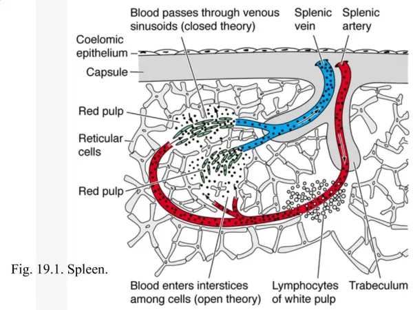
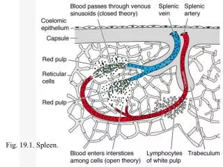
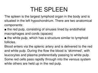
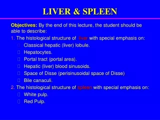
SPLENIC MICROSTRUCTURE • Fibrous framework • White pulp • Red pulp
FIBROUS FRAMEWORK • Tissue capsule • Trabeculae
CAPSULE • Continuous layer, 1.5mm thick • abundant type 1 collagen & elastin • TRABECULAE extend downwards into substance of spleen branch within it supporting framework • Largest enter the hilum • Provide a conduit for splenic A&V,n
WHITE PULP • 0.25-1mm in diameter • White semi- opaque dots on cut section • Aggregations of T lymphocytes terminations of splenic arteriolar adventitia periarteriolar lymphatic sheath ( PALS )
PALS enlarged by aggregation of B lymphocytes LYMPHOID FOLLICLES (MALPIGHIAN BODIES) • Centres of lymphocyte proliferation and differentiation antigenically stimulated GERMINAL CENTRES • Within follicles, splenic arterioles branch to form a series of parallel terminal arterioles PENICILLI
RED PULP • 75% of total splenic volume • Contain VENOUS SINUSES splenic veins • Sep. by fibrocellular network RETICULUM fibroblasts (reticular cells) & collagen(splenic macrophages)
VENOUS SINUSES • Elongated ovoid vessels 50microm. • Lined by incomplete endothelium aligned along long axis (stave cells) attached by intercellular junctions (blood passes from splenic cords to sinus lumen) luminal &external surface bears short irregular microvilli Supported circumferential &longitudinal reticular fibres
MARGINAL ZONE • At interface of red pulp and white pulp • Region where the blood is delivered to the red pulp • Lymphocytes leave the circulation to migrate to T and B lymphocyte areas of the white pulp • Aggregation of macrophages surround arteriolar ends- periarteriolar macrophage sheath (Schweigger-Seidel)
SURFACE ANATOMY • Spleen is marked on Lt. side of back with long axis along line of 10th rib • Upper border upper border of 9th rib • Lower border lower border of 11th rib • Ant. extends to mid –axillary line • Post. 4cm lateral to 10th thoracic spine
VASCULAR SUPPLY • ARTERIAL SUPPLY • Splenic A. – large,tortuous • Divides in lienorenal lig. into 2-3 main branches4-5 segmental br. • In each segment ,ramifies within trabeculae to supply parenchyma and capsule
VENOUS SUPPLY • Minor veins pass from red pulp segmental veinsdrain into main splenic V. in lienorenal lig. • LYMPHATIC SUPPLY • Drain along trabeculae • Pancreaticosplenic and coeliac nodes
SPLENIC MICROCIRCULATION • OPEN CIRCULATION • Arteriolar blood reticular spaces in splenic cords enter sinuses through minute slits • CLOSED CIRCULATION • Arteriolar blood venous sinuses
NERVE SUPPLY • Arise from coeliac sympathetic plexus • Mainly adrenergic vasomotor • Regulate of blood flow through spleen
PHYSIOLOGY • As plasma rich blood pass through sinuses: 1.filtered 2.particles phagocytosed sinus splenic pulp pressure reflects pressure throughout the portal system90% blood course through OPEN circulation
CONTD. • ERYTHROCYTE CLEARANCE • Abnormal, aged cleared • Approx. 20 ml red cells removed/day • Factors: • change in biophysical properties • hypoxic, acidotic, glucose deprived environment
fall in cellular ATP levels • loss of ATP dependent functions • sodium and calcium efflux • loss of membrane integrity