Unit 3 Trace Evidence
Unit 3 Trace Evidence. Students will identify and analyze trace evidence. Vocabulary. Compound microscope Monocular Binocular Stage Body Tube Arm Objective Nosepiece Base Lens Focus Microbe Trace evidence Wet mount slide Pollen Spore Palynology Physical property

Unit 3 Trace Evidence
E N D
Presentation Transcript
Unit 3 Trace Evidence Students will identify and analyze trace evidence
Vocabulary Compound microscope Monocular Binocular Stage Body Tube Arm Objective Nosepiece Base Lens Focus Microbe Trace evidence Wet mount slide Pollen Spore Palynology Physical property Chemical property Decant Cheiloscopy
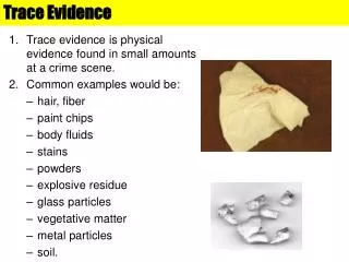
IDENTIFY TRACE EVIDENCE USING A MICROSCOPE Trace evidence is normally caused by objects or substances contacting one another, and leaving a minute sample on the contact surfaces. At a crime or accident scene, trace evidence is something that can only be detected through a deliberate processing procedure. An individual entering any environment will deposit traces of his or her presence and this material can be used as evidence. There will always be a transfer of evidence but it is very difficult to find and collect this evidence.
TRACE EVIDENCE TERMS TO KNOW Case study: http://news.bbc.co.uk/2/hi/science/nature/3640788.stm http://www.pbs.org/wnet/nature/episodes/crime-scene-creatures/case-files/297/ - read case 2
IDENTIFY TRACE EVIDENCE USING A MICROSCOPE • Types of trace evidence. • 1. Fibers • 2. Glass • 3. Hair/fur • 4. Paint • 5. Soil • 6. Impressions • 7. Gunshot residue • 8. Blood • 9. Pollen, spores • 10. And ?? List any other types of evidence which could be used as trace evidence
IDENTIFY TRACE EVIDENCE USING A MICROSCOPE Location of trace evidence. Trace can be found everywhere, but that doesn’t make it evidence. 1. Weapon a. Blood/tissue from victim b. Blood/fingerprints from suspect c. Fibers 2. Location of crime a. Blood/tissue from victim b. Blood from suspect 3. Victim a. Blood/semen from suspect b. Fibers 4. Suspect a. Blood/tissue from victim b. Fibers
IDENTIFY TRACE EVIDENCE USING A MICROSCOPE • D. Collecting trace evidence. • 1. Individuals who collect trace evidence. • A. Police Officer • B. Crime Scene Investigator • C. Forensic Scientist • 2. Trace evidence collection methods. • A. Visual inspection • (1). Use of naked eye or hand lens. • (2). Evidence removed and packaged for later analysis. • (3). Use bright light and forceps to collect evidence. • (4). Small plastic bags, glass vials, or paper using a druggist fold is used to collect evidence. Everything is double packaged and labeled.
Collecting Trace Evidence • E. Classifying trace evidence. • 1. Most trace evidence is classified using class characteristics. • A. Color • B. Shape • C. Refractive index • D. If physical properties differ then they did not come from the same source
MICROSCOPES! • A. Types of microscopes • 1. Compound microscope. • A. The eyepiece system may be monocular, binocular, or trinocular. • B. Monocular observation uses one eyepiece, binocular observation uses two. Trinocular microscopes contain an additional upright ocular which is utilized when the microscope is used with a photo or video system. • C. Compound microscopes are used to observe small animals in water, sections of plants, and/or animal and plant cells, hair & fibers
MICROSCOPES!. • B. Parts of a microscope • 1. Eyepiece (Ocular)—contains lenses to increase magnification. It may be replaced with lower or higher magnification. • 2. Body Tube—holds lenses of ocular and objectives at the proper working distance from each other. • 3. Nosepiece—permits interchange of objectives. • 4. Objectives—Contains lenses of different magnifications: usually low, medium and high power objective magnifiers. • 5. Stage—supports slide over opening that admits light from mirror or lamp.
MICROSCOPES! • 6.Stage Clips—holds slide firmly in place. • 7. Course Adjustment—moves body tube or stage up and down. • 8. Fine Adjustment—permits exact focusing by moving stage or body tube up or down very slightly. • 9. Diaphragm—regulates the amount of light passing through the specimen. • 10.Light Source—directs light upward through the diaphragm and hole in stage. • 11. Arm—supports the body tube and course adjustment. • 12. Base—firm support that bears the weight of the microscope.
MICROSCOPES! How to use a microscope 1. Carry the microscope with one hand on the arm and one hand on the base. Carry it close to the body. 2. Remove the cover and plug in the microscope. If the cord is too long, place the excess cord on the table (do not let the cord dangle over the edge of the table). 3. Always start with the lowest power. Place the slide on the microscope stage with the specimen directly over the center of the glass circle on the stage (directly over the light).
DEMONSTRATE PROPER USE, CLEANING, AND STORAGE OF A COMPOUND MICROSCOPE AND STEROSCOPE. 4. On the low power, lower the objective lens using the course (large) adjustment until it is at the lowest point. Then slowly raise the lens using the course (large) adjustment until you see the specimen within the field of view. Then use the fine (small) adjustment to see the specimen clearly. 5. At this point adjust the diaphragm while still looking through the eyepiece. This will allow more or less light. More detail will be seen when less light is allowed in through the diaphragm. Too much light will give the specimen a washed-out appearance.
DEMONSTRATE PROPER USE, CLEANING, AND STORAGE OF A COMPOUND MICROSCOPE AND STEROSCOPE. 6. Once the specimen is clear on low power, center the specimen in the field of view, then, without changing the focus knobs, switch to the next higher power. 7. Once on this higher power only the fine adjustment can be used. Use of the course adjustment at this point may scratch or crack the slide. 8. The highest power is an oil immersion lens. If this lens is used without oil, it may ruin the lens.
Identify Trace Evidence Slides Using the Trace Evidence Slide Lab Paper, fill in the table on the paper, being sure to add as much detail to your drawings and descriptions as possible. You need to have an example for each of the 19 different trace evidence slides. Your quiz will consist of ten slides set up on a microscope with multiple choice answers.
Collect Trace Evidence Using the packing tape provided, obtain two samples of “trace evidence,” one from your shirt and one from the bottom of your shoe. 1. Obtain a 2-3 inch length of clear packing tape, being careful to only touch the very edge of the tape. 2. Press the tape to your shirt/bottom of shoe 3. Carefully peel away the tape and place it sticky side down on the transparency. 4. Using a microscope, make 6 (3 for each sample) observational drawings of what you see. 5. Identify as many particles as you can (at least 10 per sample)
Collecting Trace Evidence • b. Tape lift • (1). Clear tape is used. • (2). Repeatedly apply tape to small area until most of the stickiness is gone. • (3). Tape is folded back upon itself, taped to a glass slide or taped to a piece of plastic. • c. Vacuum • (1). Nozzle should be short and transparent. • (2). Debris is collected on a filter or membrane.
MICROSCOPES! • a. Trouble shooting. • (1). If you wear glasses take them off. • (2). If you see only your eyelashes, move closer. • (3). If using a monocular microscope, cover or close your other eye. • (4). If you see a dark line that goes part way across the field of view, turn the eyepiece. The dark line is a pointer that is a very valuable tool when you want to point out something to your lab partner or your teacher.
DEMONSTRATE PROPER USE, CLEANING, AND STORAGE OF A COMPOUND MICROSCOPE AND STEROSCOPE. • How to clean and store a microscope • 1. Only use lens paper to clean the lenses. A paper towel may be used to clean the stage. • 2. Lower the stage completely. • 3. Return the lens to the lowest power. • 4. Wrap cords loosely around the microscope and replace the cover. • 5. Return the microscope to the storage area carrying the microscope with one hand on the arm and one hand on the base and close to the body.
3.10 PREPARE A WET MOUNT SLIDE • A. Wet mount slide preparation. • 1. Use a flat glass slide. The slide should be clean and free of dust or other particles. • 2. Draw a few drops of the liquid specimen into a medicine dropper. • 3. Pick up the glass slide with one hand grasping it by the outer edges. • 4. Place one drop of the liquid specimen from the medicine dropper on top of the slide. The drop should be placed in the center of the slide. • 5. Do not tilt the slide in your hand.
PREPARE A WET MOUNT SLIDE • 6. Use your free hand to carefully pick up a cover slip (they are extremely fragile) grasping it by the outer edges. • 7. On an angle, place the cover slip on top of the specimen on the slide. The edges of the cover slip should match up with the edges of the slide. DO NOT PRESS DOWN ON THE COVER SLIP. • 8. Holding the slide by the outer edges, keep the slide as horizontal and steady as possible. Place the slide on the stage of the microscope. The specimen should be placed over the opening of the diaphragm.
IDENTIFY MICROBES • IDENTIFY MICROBES USING MEASUREMENT AND MICROSCOPY TECHNIQUES IN A SIMULATED PROFESSIONAL SETTING. • A. Microbial forensics uses molecular techniques to analyze microscopic microbes that cause illness or death (i.e., bacteria, viruses, and fungi as well as any pathogenic agent that can be used as a biological weapon). Microbial forensics includes the full scope of forensic evidence, such as analysis of microbes, materials used to prepare, stabilize, and deliver the toxin or pathogen, and fingerprints, hair, fiber, and pollen. • B. Laboratory analysis used in microbial forensics including molecular sequencing, microbiological cultures, biochemistry, electron microscopy, crystallography, and mass spectrometry. • Watch the video “Classifying Monerans and Protists” from the WHS catalog. Search for “protists.” Complete the accompanying handout.
Microbe Murder Complete the lab as described
Sealed with a Kiss Lab Chromatography: using a solvent to separate a mixture.
Analysis of White Powders Lab Forensics textbook page 208
Labs • In this unit you should have completed the following lab assessments: • Trace evidence prepared slides • Personal trace evidence • Microbe Murder with a wet mount slide • Sealed with a Kiss – chromatography • Analysis of white powders (pg 208)
Case Study Write a case study focusing on crimes where trace evidence played a major role in convincing a jury of guilt. Keep it short – about half a page! Email it to me when finished. Critiques are starting!!!
TEST REVIEW 1. When you use paper chromatography, what is the mobile phase? What is the stationary phase? 2. What is chromatography? 3. Why is pencil and not pen used to mark the chromatography strips? 4. In the qualitative analysis of white powders, what is meant by solubility? What factors may affect the apparent solubility of the first 10 samples? Is solubility a physical or chemical property?
TEST REVIEW 5. In the above mentioned white powders lab, what indicates that a chemical reaction has taken place? What is Rf, and how is it calculated? Use the internet to determine whether lip prints have been used as evidence in a trial. Is this related to the Daubert Ruling? Discuss