Unit 4: Trace Evidence ( Chapter 16)
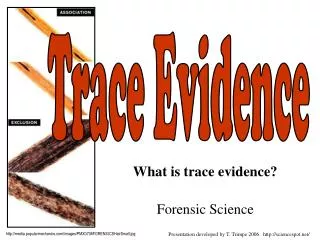
Unit 4: Trace Evidence ( Chapter 16). I. Trace evidence=. Physical evidence found at a crime scene in small but measurable amounts Examples: hair, glass, fibers, paint, pollen, gunshot residue, soil, etc. II. Examination of Trace Evidence uses Microanalysis.

Unit 4: Trace Evidence ( Chapter 16)
E N D
Presentation Transcript
I. Trace evidence= • Physical evidence found at a crime scene in small but measurable amounts • Examples: hair, glass, fibers, paint, pollen, gunshot residue, soil, etc.
II. Examination of Trace Evidence uses Microanalysis • Microanalysis= application of a microscope to the observation, collection, and analysis of trace evidence • Purpose= To determine if an association of persons, places, and things can be made AND the strength of that association
III. Instruments of Microanalysis • 1. Light microscopes • Total magnification = eyepiece mag. (x) objective mag. TM = EP (x) OBJ • Amount of detail seen/revealed is related to the resolving power of the microscope . . .which is the ability to distinguish differences in fine structure.
1. Light microscopes cont. • Types of light microscopes • A. Stereo binocular microscope • Used in preliminary evaluation of trace evidence • Constructed fro 2 separate optical microscopes for observation of each eye simultaneously • Utilize reflected light • View has 3-D appearance
1. Light microscopes cont. • Types of light microscopes • B. Compound light microscope • Most often employed with transmitted bright-field illumination • Allows to see details of construction of evidence Amoeba magnified 100 x
1. Light microscopes cont. • Types of light microscopes • B. Polarized light microscope • Attachment to light microscope that allows for plain polarized light • Vibrations of normal light are in all directions (360 degrees), but if restrict vibration to one direction, you get plain polarized light • Can give better quantitative data (measurements can be made more easily)
Images from: http://www.microscopyu.com/articles/polarized/polarizedintro.html
2. Scanning Electron microscope (SEM) • beams of electrons bounced off surface of specimen to produce image • can magnify much more than light microscope . . . SEM has possible range of 10x to 100,000 x Try it! Ant magnified ~1500 x
3. Microspectrophotometry • Combines microscope with a spectrophotometer so that light absorption properties of a very small sample can be recorded • Helps experts to determine COLOR of a sample. . . such as with fibers, hair, and painted surfaces
Part A e
Part B Illustrates: Depth of field -Or- Depth of focus